Raman Microscopic Analysis of Grain Kernels
A new Raman Video Microscope Sampling System has been developed for precision sampling during Raman spectroscopy analysis. The Raman analysis of grain kernels using this system reveals distinct Raman shifts, which would otherwise be washed out by a strong fluorescence background.
B&W Tek's new product — Raman Video Microscope Sampling System, is a video microscope with integrated camera and LED illuminator to allow precision spot sampling. Focusing and positioning of the excitation laser beam is facilitated by the video camera which provides imaged details of the sampling spots. The sampling image can be easily recorded and saved, as well as included in the test report together with measured spectra through BWSpec™ software, which is the operational software for all B&W Tek Raman spectrometers. The system provides both bright field and dark field illuminations to optimize the illumination required for various sample surfaces. It is compatible with B&W Tek's complete line of laboratory and industrial Raman probes. When coupled with B&W Tek's portable Raman spectrometers, this video microscope system provides the advantages of a Raman microscopy at a fraction of the cost of most research instruments.
The application discussed in this paper is the Raman analysis of a grain kernel. Without the Video Microscope Sampling System, the Raman signal is overwhelmed by the strong fluorescence background generated from the grain bran (thickness 60 ~ 70 μm) (1). The Raman analysis of a sliced cross section of the kernel under the Video Microscope Sampling System enables the identification of specific components, such as starch and lipids, inside the kernel.
Experiment and Results
The experimental setup is shown in Figure 1(a). The Raman analysis was done using B&W Tek's MiniRam™ Spectrometer with a 785 nm excitation laser. The laser spot size is about 90 μm in diameter with 20× objective lens. Samples were prepared by polishing a cross section of the grain to generate a surface suitable for microscopic imaging. The microscope image captured through BWSpec™ is shown in Figure 1(b).

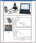
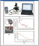
Figure 1: (a) Experimental setup. (b) Grain kernel microscopic image captured through BWSpecâ¢. (c) Comparison of Raman spectrum of a grain kernel using B&W Teks MiniRam⢠under Video Microscope Sampling System (upper) and a Raman spectrum of a whole grain kernel using B&W Teks MiniRam⢠but without using Video Microscope Sampling System (lower).
The Raman analysis results are shown in Figure 1(c). Raman analysis of the cross section of the grain kernel using the Video Microscope Sampling System shows distinct Raman shifts associated with starch and lipids, while the analysis on a whole grain kernel without Video Microscope Sampling System results in Raman peaks washed out by the fluorescence background generated from the grain bran.
Conclusions
B&W Tek's Video Microscope Sampling System, when coupled with B&W Tek's Raman spectrometers, can direct laser excitation on the specific area or specific structure of the sample, which provides the advantages of choosing an area to be analyzed, or avoiding areas so as to minimize fluorescence.
Reference
(1) K. Kulp and J.G. Ponte, Handbook of Cereal Science and Technology (CRC Press, 2nd edition, 2000), p. 2.


B&W Tek, Inc.
19 Shea Way
Newark, DE 19713
Tel. 302-368-7824, Fax 302-368-7830
Email: info@bwtek.com
Website: www.bwtek.com


The World of Microplastics Up to Date – an Overview
April 23rd 2024Watch this 20-minute educational video by Andreas Kerstan, Agilent Product Specialist in molecular spectroscopy, to gain a comprehensive update on the microplastics landscape and the environmental concerns related to them. Discover the current challenges in microplastics characterization and how Agilent innovative solutions and techniques, including FTIR, LDIR, GC/MS, and ICP-MS, are addressing these issues head-on.
Accurate Microplastics Analysis in Minutes, Not Hours
April 10th 2024The automated Agilent 8700 LDIR chemical imaging system lets you obtain high-quality images and spectral data faster than ever before. So, you can perform confident large-scale microplastics studies and monitoring activities.

