Long-Wavelength Dispersive 1064nm Raman: Non-Invasive Cancer Tissue Diagnostics
Overcoming fluorescence background noise in cancerous biological tissue samples achieved by using Dispersive 1064nm Raman excitation instruments.
It has been documented that Raman spectroscopy is an effective technique for detecting molecular changes associated with cancer; however, it is well-known that fluorescence found in biological samples generates significant noise complicating specificity in distinguishing between normal and cancerous tissue (1).
Raman scattering is a spectroscopic technique capable of providing highly detailed chemical information about a tissue sample. In contrast to other optical spectroscopic techniques, there are a large number of Raman-active molecules in tissue and their spectral signatures are sharp and well delineated.

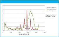
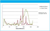
Figure 1: Raman spectra of normal human kidney tissue and cancerous tissue.
The earliest Raman spectroscopy techniques utilized Fourier transform (FT)–Raman spectroscopy, a method that measures Raman spectra with high signal-to-noise ratio (S/N) and minimal fluorescence interference and has been used for many in vitro applications. However, long integration times and bulky instrumentation negate this technique for in vivo use.
Early attempts at measuring Raman spectra of tissue were difficult because of the fluorescent nature of tissue and limitations of sources and detectors. Today, because of the telecom boom and bust, there has been a Manhattan Project effect on the development of lasers and optical components. Ten years ago, optics, lasers, and detectors were limited to laboratories on bulky optical benches with sophisticated liquid nitrogen cooling and complicated alignment/operations requiring a PhD to operate.
Current chemometric methods rely on mathematical techniques such as the use of second derivatives, Fourier filtering, and polynomial fitting to remove the fluorescence background. Classification algorithms are developed using a variety of multivariate statistical methods such as linear and nonlinear discrimination analysis, neural networks, genetic algorithms, and cluster analysis. The resulting end goal is to obtain high sensitivity and specificity in the recognition of a target condition amidst a variety of tissue categories depending upon the tissue type.
Today, without sacrificing performance in many cases, lasers are the size of your little finger; Newton's prism has been replaced by a high-throughput holographic volume phase grating; and detectors can be thermoelectrically air-cooled, doing away with liquid nitrogen. Using low-profile, surface-mount electronics completes the picture, enabling spectrometers the size of your Blackberry or iPhone.
Therefore, for the first time, a ruggedized no moving parts dispersive long-wavelength 1064 nm Raman spectroscopy in conjunction with multivariate statistical technique has the potential for rapid in vivo diagnosis of benign and malignant tissues based on the optical evaluation of spectral features of biomolecules.
For more information contact us at info@bayspec.com.


BaySpec, Inc.
1101 McKay Drive, San Jose, CA 95131
tel. (408) 512-5928
Website: www.bayspec.com
Email: info@bayspec.com
References
(1) A. Mahadevan-Jansen, Biomedical Photonics Handbook, T. Vo-Dinh, Ed. (CRC Press, Washington, D.C., 2003), pp. 30:1– 30:27.
(2) Charge-Transfer Devices in Spectroscopy, J.V. Sweedler (Editor), Kenneth L. Ratzlaff (Editor), B.M. Denton (Editor), published by John Wiley & Sons, Inc., March 1994.


Single Cell and Microplastic Analysis by ICP-MS with Automated Micro-Flow Sample Introduction
April 25th 2024Single cell ICP-MS (scICP-MS) is increasingly seen as a powerful and fast tool for the measurement of elements in individual cells, mainly due to the high sensitivity and selectivity of ICP-MS. Analysis is performed in the same way as single nanoparticle (spICP-MS) analysis, which has become a well-established technique for the analysis of nanoparticles and particles.
Hot News on Agilent LDIR, New Developments, and Future Perspective
April 25th 2024Watch this video featuring Darren Robey and Dr. Wesam Alwan from Agilent Technologies to gain insights into the future trends shaping microplastics research and the challenges of their characterization. Discover the essential components necessary for accurate microplastics analysis and learn how the Agilent 8700 LDIR system addresses these challenges. Offering rapid and precise analysis capabilities, along with easy sample preparation methods that minimize contamination, the Agilent 8700 LDIR system is at the forefront of advancing microplastics research.
The World of Microplastics Up to Date – an Overview
April 23rd 2024Watch this 20-minute educational video by Andreas Kerstan, Agilent Product Specialist in molecular spectroscopy, to gain a comprehensive update on the microplastics landscape and the environmental concerns related to them. Discover the current challenges in microplastics characterization and how Agilent innovative solutions and techniques, including FTIR, LDIR, GC/MS, and ICP-MS, are addressing these issues head-on.

