Measuring Cellular-Scale Nutrient Distribution in Algal Biofilms with Synchrotron Confocal Infrared Microspectroscopy
The authors use synchrotron confocal IMS to measure the response of two dominant algal species from a stream of biofilm to a reduction in nutrient availability.
The microscope and infrared spectrometer are two of the most useful tools for the study of biological materials, and their combined analytical power far exceeds the sum of the two. Performing molecular spectroscopy through a microscope superimposes chemical information onto the physical microstructure obtained from the optical microscope when visible and infrared information are collected under the same conditions. The instrument developments that enable current infrared microspectroscopic studies began with the introduction of the first research-grade infrared microscope, patented in 1989 (1). By 1993, published reports using this method to determine macroalgae (seaweed) cell-wall composition appeared (2-4). Since these initial reports, the use of infrared microspectroscopy (IMS) in microalgal (single cells or groups of cells) research has grown. Primarily, cultured algae have been used to hone IMS methodology and evaluate its capabilities in algal research (5–8). Studies involving natural, mixed species assemblages, which can utilize the spatial resolution potential of this technique fully are rare (9–11). For instance, in a recent review of IMS microalgal ecological research (12), only 3 of the 29 peer-reviewed publications investigated natural algal assemblages. Both thermal and synchrotron infrared sources provide a resolution capable of measuring individual algae in mixed species assemblages, and each has its advantages. For example, thermal source IMS is more accessible, allowing more samples to be analyzed than synchrotron IMS. However, synchrotron IMS with confocal masking provides superior resolution, which can be critical in isolating small or contiguous cells.
Algal ecology is the study of the interaction between algae and their environment. Infrared microspectroscopy addresses a major logistical problem in this field, obtaining species-specific cellular biochemical information from natural, mixed-species assemblages (11,12). Benthic (bottom-dwelling) algae, for example, grow in a three-dimensional matrix (biofilm) composed of different cell sizes, shapes, and configurations. The optical and ecological challenge of studying algae is apparent from Figure 1, which shows a photomicrograph of algal chlorophyll fluorescence on a rock. Several issues make it difficult to obtain single species measurements with standard techniques: cell sizes can vary over an order of magnitude; species can occur as single cells, long filaments, or globular colonies; a number of different species can be found within a few square millimeters; and fluorescence can vary across cells (that is, the physiological state varies across cells).

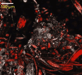

Figure 1: Micrograph of algal chlorophyll fluorescence (red) on a rock (white) collected with confocal laser scanning microscopy. Note the variety of cell shape configurations and sizes among the benthic algal species. Larger species and colonies are routinely analyzed by conventional IMS. The small size of many species (


Getting accurate IR spectra on monolayer of molecules
April 18th 2024Creating uniform and repeatable monolayers is incredibly important for both scientific pursuits as well as the manufacturing of products in semiconductor, biotechnology, and. other industries. However, measuring monolayers and functionalized surfaces directly is. difficult, and many rely on a variety of characterization techniques that when used together can provide some degree of confidence. By combining non-contact atomic force microscopy (AFM) and IR spectroscopy, IR PiFM provides sensitive and accurate analysis of sub-monolayer of molecules without the concern of tip-sample cross contamination. Dr. Sung Park, Molecular Vista, joined Spectroscopy to provide insights on how IR PiFM can acquire IR signature of monolayer films due to its unique implementation.


Deep Level Transient Spectroscopy Reveals Influence of Defects on 2D Semiconductor Devices
April 25th 2024A recent study used deep level transient spectroscopy to investigate the electrical response of defect filling and emission in monolayer metal-organic chemical vapor deposition (MOCVD)-grown materials deposited on complementary metal-oxide-semiconductor (CMOS)-compatible substrates.

