|Articles|May 13, 2019
Raman Advances Using SESORRS and SERS for Biomedical Measurements
Author(s)Jerome Workman, Jr.
Recent advances in Raman spectroscopy, specifically using surface enhanced spatially offset resonance Raman spectroscopy (SESORRS), which is a combination of surface enhanced Raman scattering (SERS), and spatially offset Raman spectroscopy (SORS) are enabling noninvasive, real-time measurements of living tissue and multiple bacterial pathogens. In an interview with Karen Faulds, the 2019 recipient of the FACSS Charles Mann Award for Applied Raman Spectroscopy, we explore the latest developments in Raman spectroscopy for biomedical analysis applications. This interview is part of a series of interviews with the winners of awards presented at the SciX conference.
Advertisement
Recent advances in Raman spectroscopy, specifically using surface enhanced spatially offset resonance Raman spectroscopy (SESORRS), which is a combination of surface enhanced Raman scattering (SERS), and spatially offset Raman spectroscopy (SORS) are enabling noninvasive, real-time measurements of living tissue and multiple bacterial pathogens. In an interview with Karen Faulds, the 2019 recipient of the FACSS Charles Mann Award for Applied Raman Spectroscopy, we explore the latest developments in Raman spectroscopy for biomedical analysis applications. This interview is part of a series of interviews with the winners of awards presented at the SciX conference.
You have published research using handheld surface enhanced spatially offset resonance Raman spectroscopy (SESORRS) for noninvasive measurements of live breast cancer tumor cells (1). What prompted you to investigate this specific problem? What are the unique aspects of your measurement approach?
We are very interested in being able to carry out Raman measurements at depth with a long term view that this approach could one day be used in vivo. My close collaborator at Strathclyde, Duncan Graham, and I published the first papers on the combination of spatially offset Raman spectroscopy (SORS) and surface enhanced Raman scattering (SERS), or SESORS, in collaboration with Nick Stone at the University of Exeter, and Pavel Matousek from Rutherford Appleton Laboratory, in 2010–2011 (2,3). This demonstrated the detection of nanotags buried at depths of 25 mm in tissue. Our recent work carried out by PhD student Fay Nicolson has extended this to use the resonance effect, where we use Raman reporters that are in resonance with the SORS excitation wavelength of 830 nm, to further enhance the signal to give SESORRS (a bit of a mouthful!). This allowed detection of the resonant nanotags at depths of 25 mm using a handheld instrument (the initial work was carried out using a benchtop instrument). We also extended the work to the detection of the resonant nanotags inside a cancer tumor model buried at depth. We grew 3D multicellular tumor spheroids (MTS) from breast cancer cells, these cell models resemble the 3D in vivo environment of a tumor more closely than 2D models, establishing characteristic concentration gradients in oxygen, nutrients, and metabolites. Our resonant nanotags were up taken into the spheroid model and we were able to detect these at depths of up to 15 mm. This is the first report of the detection of nanoparticles using SESORRS within a tumor model which has potential applications for the noninvasive detection of breast cancer using targeted nanoparticles combined with optical detection.
You have also recently developed a new bionanosensor for the isolation and detection of multiple bacterial pathogens via magnetic separation and surface enhanced Raman scattering (SERS) for analysis of multiple types of antimicrobial-resistant pathogens (4). How did you develop this approach? What unique discoveries did you find in developing this technique?
The main aim of this approach was to develop a method for the detection of bacteria in complex samples, for example, in clinical or food samples. We wanted to develop an approach that could be used to both isolate the bacteria, allowing it to be separated from the complex matrix, and then to detect the bacteria using the same bionanosensor. Our postdoc, Hayleigh Kearns, was able to do this by making magnetic nanoparticles for isolation and separation which we then functionalized with lectins which would non-specifically capture multiple bacteria. We then developed SERS nanotags that had specific Raman reporters and antibodies that could then be used to specifically identify the bacteria strains present. This allowed multiple bacteria to be detected in one sample, using one bionanosensor and one measurement very sensitively using portable detection. We have been collaborating with Roy Goodacre of the University of Liverpool on the deconvolution of multiplexed SERS signals and this has been an important step forward allowing us to both multiplex and quantify the components of a multiplex.
Would you explain the use of red-shifted chalcogen pyrylium-based Raman reporters in terms of their chemical properties and how they are used?
The chalcogen dyes are made by our collaborator, Professor Mike Detty from the University at Buffalo, New York. These are excellent molecules which bind strongly to the metal surface to give a strong SERS response. However, their main advantage is that Mike can tune the molecules to absorb at NIR wavelengths. If we can tune the absorbance of the dyes to the laser excitation wavelength then we can get excellent resonant enhancement from these wonderful molecules. There are not many nonfluorescent, NIR absorbing dyes commercially available that attach strongly to metal surfaces so these molecules make excellent resonant Raman reporters that give strong SERS in the NIR biological window which is of interest for in vivo measurements.
What instrumentation or sampling advances did you have to develop in order to perform your research?
We have only used commercially available instrumentation for this work although we do have a homemade SORS system. The handheld SORS instrument is commercially available, and we are working with another company on the bacteria detection work. Fay developed a set up that allowed the SORS instrument to be mounted above a microscope stage that allowed her to carry out crude imaging using the handheld instrument. The advantages of both these instruments is their portability which makes them very attractive for use for in situ measurements, whether in the clinic or the food manufacturing environment.
From your perspective what are the most exciting developments in Raman spectroscopy for biomedical analysis over the past five years (in terms of both applications and instrumentation development)?
In terms of instrumentation, the development of portable instruments is of great interest. Small, portable instruments are now available which have excellent performance and are not compromised in terms of specification compared to some benchtop instruments. These are ideal for the type of aim and point, rapid measurements we are interested in for some of these in situ measurements. In terms of application, there is some really exciting work going on using Raman tags which work in the cell silent region of the spectrum (1800–2800 cm-1) for imaging applications. This area of research was first published by Katsumasa Fujita and colleagues from Osaka University who used Raman combined with alkyne molecules to shift the bands into this cell silent region. He is also doing some excellent work on increasing the speed of Raman imaging to make acquisition of spectral maps much faster. Wei Min of Columbia University is now doing some really nice work using these biorthogonal labels using stimulated Raman spectroscopy (SRS) and this is a really exciting area of research that I think will become really important in biomedical imaging in the future.
What have been your greatest challenges in scientific discovery over your career? What is your general approach to problem solving in your scientific work?
One of the greatest challenges in SERS is making the measurements reproducible and quantitative as well as the complexities in developing multiplexed SERS assays and interpreting the data. We generally have a good handle on this, however there is a lot of work and problem solving that goes on by the researchers in our research group to achieve this, the outside world see the final work in publications and presentations, but it often takes a lot of work to get there! It requires a lot of problem solving that involves working out what effect every component has on the final system. Multiplexed assays are very challenging to develop!
What are some major gaps in knowledge for Raman technology that you would like to see more research and development time devoted to?
Raman technology has developed exponentially in the last 10–15 years and has reduced dramatically in size and costs from when I first started doing Raman during my PhD. I think perhaps one of the areas that will be further developed over the next few years is further decreases in instrument size and the performance of these small instruments. The cost needs to decrease before it can be used in some applications, for example, in food testing as this can be prohibitive but I guess it is a bit of the-chicken-or-the-egg dilemma; if high demand for small portable instruments exists then the cost of production of instruments should decrease. I think more development could go into the front-end sampling geometries of instruments; in portable commercial instruments these tend to be fixed and less adaptable and they are not always ideal for the sample or assay the instrument is being used for in terms of beam size and geometry and sample integration. Being able to integrate bespoke data analysis directly into instruments will also be important in the future, particularly for handheld instruments where you may want to do testing in the field.
What do you anticipate is your next major area of research or application in your field?
We are extremely interested in extending the SESORRS approach further into biomedical applications and moving towards using this approach to specifically detect and target disease states with a long term view to be able to do this in vivo one day.
References
- F. Nicolson, L.E. Jamieson, S. Mabbott, K. Plakas, N.C. Shand, M.R. Detty, D. Graham, and K. Faulds, Chem. Science9(15), 3788–3792 (2018).
- N. Stone, K. Faulds, D. Graham, and P. Matousek, Anal. Chem.82(10), 3969–3973 (2010).
- N. Stone, M. Kerssens, G.R. Lloyd, K. Faulds, D. Graham, and P. Matousek, Chem. Science2(4), 776–780 (2011).
- H. Kearns, R. Goodacre, L.E. Jamieson, D. Graham, and K. Faulds, Anal. Chem.89(23), 12666–12673 (2017).
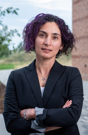
Karen Faulds is a Professor in the Department of Pure and Applied Chemistry at the University of Strathclyde and an expert in the development of surface enhanced Raman scattering (SERS) and other spectroscopic techniques for novel analytical detection strategies and in particular multiplexed bioanalytical applications. She has published over 130 peer reviewed publications and has filed five patents. She has been awarded over £20M in funding as principal and co-investigator from EPSRC, BBSRC, charities, industry and governmental bodies. Her group’s research has been recognized through multiple awards including the Nexxus Young Life Scientist of the Year award (2009), Royal Society of Chemistry (RSC) Joseph Black Award (2013), Craver Award (2016), and Charles Mann Award (2019). She is a Fellow of the RSC (2012), the Society for Applied Spectroscopy (2017) and the Royal Society of Edinburgh (2018). She has given over 75 invited talks at national and international conferences. She is the Strathclyde Director of the Centre for Doctoral Training in Optical Medical Imaging, serves on the editorial board of RSC Advances and the editorial advisory board for Analytical Chemistry, Analyst and Chemical Society Reviews and is the current Chair of the UK’s Infrared and Raman Discussion Group (IRDG).
Newsletter
Get essential updates on the latest spectroscopy technologies, regulatory standards, and best practices—subscribe today to Spectroscopy.
Advertisement


Advertisement
Advertisement