
|Articles|June 1, 2016
- Special Issues-06-01-2016
- Volume 31
- Issue 6
Using SERS to Study How Cells Respond to Pharmaceuticals
Colin Campbell discusses his work applying SERS to biomedical applications.
Advertisement
The application of surface-enhanced Raman spectroscopy (SERS) to biomedical studies is of particular interest. Colin Campbell of the University of Edinburgh is using SERS to make spatially resolved measurements in live three-dimensional (3D) cell cultures to determine the response to drugs during drug discovery operations. For this work, Campbell received a FACSS Innovation Award at the SciX 2015 conference last October. This interview is part of the SpectroscopyâSciX interview series.
An important aspect of your work using SERS in drug discovery is that you are measuring cell response within 3D cell structures. Your method involves the use of SERS nanosensors, which are incorporated into the 3D structures. Can you briefly describe the nanosensors?
Our nanosensors are gold nanoparticles coated in reporter molecules that change their structure in response to changes in their environment (pH and redox potential). The gold nanoparticles allow us to measure surface-enhanced Raman spectra of the reporters and thus the spectra that we measure give useful information on the pH and redox potential of the cells. The pH and redox potential can give us information about the type of metabolic pathway that the cells are using and thus we can get metabolic information in real-time.
After the nanostructures are taken up by the cells, how do you know where a nanosensor is at any given moment?
We can make a Raman map of a cell and find the exact location of the nanosensor. This involves measuring multiple Raman spectra across the cell and making an image based on the Raman spectra.
What are you currently using this technique to investigate?
We’re looking at the response of cells in 3D culture to perturbations such as treatment with drugs. We are seeing some of the interesting metabolic properties that we expect from 3D cultures and hope to see differences between different types of treatment.
How do those results compare to what one sees using current techniques?
Currently there is no other way to make these particular measurements in 3D culture so it’s difficult to compare. I think it’s a great strength of our technique that it gives us information that isn’t accessible otherwise. It also makes for a lot of exciting challenges for my students and me.
To read the full interview please visit:
Articles in this issue
over 9 years ago
Evaluation of Eye Shadow Compounds Using Raman Microspectroscopyover 9 years ago
Tech Tip: Choosing Raman Excitation WavelengthsNewsletter
Get essential updates on the latest spectroscopy technologies, regulatory standards, and best practices—subscribe today to Spectroscopy.
Advertisement
Related Content
Advertisement


Advertisement
Advertisement
Trending on Spectroscopy Online
1
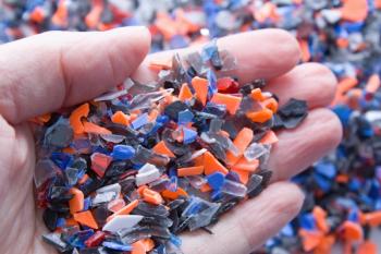
Deep Learning Meets Spectroscopy to Transform Plastic Recycling Accuracy
2
Developing LIBS for Molten Salt Reactor Monitoring
3
Previewing a Talk on Glow Discharge Optical Emission Spectroscopy
4
Ep. 42: Did You Look at the Raw Data?
5