Lipid Droplets and Endoplasmic Reticulum Analyzed Using Fluorescence Probe
Scientists from the University of Jinan developed a new fluorescent probe to better understand the relationship between certain lipids.
Scientists from the University of Jinan are looking to gain a better understanding of the relationship between lipid droplets (LDs) and endoplasmic reticulum (ER) by creating a novel polarity-sensitive
The scientists, whose work was published in Spectrochimica Acta Part A: Molecular and Biomolecular Spectroscopy, created a system based on fluorescent analysis, which is a non-invasive, fluorescence-imaging technique that provides real-time in situ visualization of activities in living cells. Using this as a basis, they created a probe LP system to be novel and polarity-sensitive, using bearings of bearing of 3-ketone-4-hydroxy-7-(diethylamino) coumarin and N, N-diethylaniline as a fluorescent reporter with a π bridge.
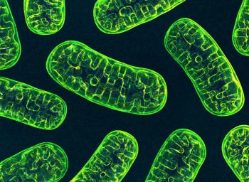
Eukaryotic cells contain many organelles, each of which have their own unique forms and functions (1). LDs are spherical organelles that have been confirmed to be vital in maintaining lipid homeostasis in cells, all while participating in vital physiological activities, such as immune responses and signal transductions. LDs have also been showed to bear strong connections to other organelles, most notably ER; in fact, it is widely believed that LD generations originate from ER. This has led to discussions on the detailed mechanisms of LDs and ER in cellular activities, leading to studies like this one.
The probe LP showed well red-shifted emissions and an increase fraction of water in 1,4-dioxane due to the intramolecular charge transfer (ICT) process. As for biological imaging, the probe LP visualized LDs and ER with both green and red fluorescence separately. The system achieved the dynamic behaviors of LDs and ER using LPduring oleic acids and starvation stimulations. “Probe LP is a valuable molecular tool for investigating the relationships of LDs and ER in various cellular activities,” the scientists said in the study (1).
Reference
(1) Dai, X.; Wang, B.; Tian, M.; Wang, J.; Dong, B.; Kong, X. Development of a high polarity-sensitive fluorescent probe for visualizing the lipid droplets and endoplasmic reticulum with dual colors in living cells. Spectrochim. Acta A Mol. Biomol. Spectrosc. 2023, 301, 122973. DOI:
Newsletter
Get essential updates on the latest spectroscopy technologies, regulatory standards, and best practices—subscribe today to Spectroscopy.


![Figure 3: Plots of lg[(F0-F)/F] vs. lg[Q] of ZNF191(243-368) by DNA.](https://cdn.sanity.io/images/0vv8moc6/spectroscopy/a1aa032a5c8b165ac1a84e997ece7c4311d5322d-620x432.png?w=350&fit=crop&auto=format)

