



Surface-enhanced Raman spectroscopy (SERS)
Latest News
Advertisement
Latest Videos




Shorts

Advertisement
More News

Wednesday's program at SciX 2026 kept returning to one question: can spectroscopy perform outside the controlled laboratory? Speakers took surface-enhanced Raman scattering (SERS), laser-induced breakdown spectroscopy (LIBS), mid-infrared photonics, chemometrics, and fluorescence excitation–emission matrix (EEM) spectroscopy into clinics, food plants, reactor coolant loops, brine fields, and drinking water supplies.
Raman spectroscopy is entering one of its most consequential periods since its commercialization, driven by artificial intelligence, coherent heterodyne detection, and radical instrument miniaturization, with new deep-learning frameworks and portable systems moving Raman analysis into fields, factories, and crime scenes.
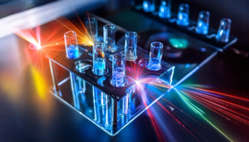
Raman spectroscopy just got a brain, a stopwatch, and a nose. Together, these upgrades are turning a century-old light-scattering trick into a frontline tool for catching cancer earlier, chiral drugs cleaner, and toxic chemicals faster than ever before.
Reliability, dynamic measurements, artificial intelligence, and real-world applications are reshaping Raman spectroscopy – while Molecular Photonics points toward new translational frontiers.
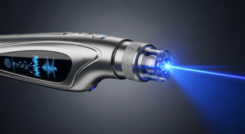
Raman spectroscopy is shedding its bulky, benchtop reputation as chip-scale spectrometers, tip-enhanced probes, and AI-native detectors push the technique into pockets, production lines, and single molecules. The result is an instrument category being rebuilt from the optics up, faster, smaller, deeper, and smarter than the Raman systems of even five years ago.

Researchers developed spherical and hollow gold nanoparticle-based SERS platforms that enable label-free molecular imaging of chemotherapy drug effects and drug release within cancer cells, offering a theranostic tool with potential applications in early cancer diagnostics.

Measuring neonicotinoids in drinking water is not heavily regulated in the United States, but low-cost, rapid analytical tools are changing the conversation.

A recent study examined how surface-enhanced Raman spectroscopy (SERS) is being used with machine learning (ML) analysis to accurately distinguish patients with Crohn’s disease.
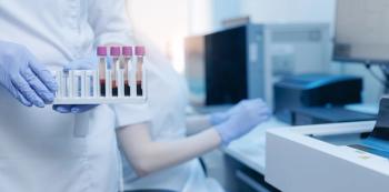
A new diagnostic platform may help detect drug-induced liver injury better. How would this work in real-world clinical settings?
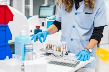
The combination of surface-enhanced Raman spectroscopy (SERS) and digital microfluidics (DMF) might be able to improve detection of liver injury biomarkers.

A recent study showcased a wearable, glove-mounted sensor that can detect trace residues of two cancer drugs on workplace surfaces.

In this interview clip, Sian Sloan-Dennison, a postdoctoral research associate at the University of Strathclyde, discusses how a new digital microfluidics surface-enhanced Raman spectroscopy (DMF-SERS) platform be implemented in clinical settings.
Why is determining the function of a surface-enhanced Raman spectroscopy (SERS) assay important? This "Pathways in Spectroscopy" clip offers career advice for researchers in biological and clinical analysis by addressing this question.
Why does integrating a surface-enhanced Raman scattering (SERS) assay with a digital microfluidics (DMF) platform improve sensitivity, reproducibility, and throughput when detecting onset of drug-induced liver injury?
An upcoming two-part video series on “Pathways in Spectroscopy” tackles working with surface-enhanced Raman spectroscopy (SERS), offering practical advice for researchers in clinical laboratories.
Advertisement
Advertisement
Trending on Spectroscopy Online
1
Quantitative Raman Spectroscopy and the Unsolved Puzzle of Spectral Interpretation
2
Thermo Fisher Introduces Benchtop Spark-OES Analyzer for Routine Metals Quality Control
3
IRDG Christmas Meeting to Spotlight Biomedical Vibrational Spectroscopy in London
4
Curated 2026 Evergreen Content from Spectroscopy
5