





Biological, Medical, and Clinical Analysis
Latest News
Advertisement
Latest Videos





Shorts
0:43
How Surgeons Can Use Raman Probes
4 days ago
by
Jürgen Popp
1:21
Winning the 2026 Ellis R. Lippincott Award
a month ago
by
Jürgen Popp
0:52
Translating Chemical Bond Imaging Toward Disease Diagnosis
a month ago
by
Ji-Xin Cheng
0:31
Best of the Week: Implementing SERS in Clinical Settings, How Lasers Became Valuable Tools in Spectroscopy
6 months ago
by
Will Wetzel
Advertisement
More News

Atomic spectroscopy has quietly gone from counting elements in bulk to counting single nanoparticles, imaging tissue at the micrometer scale, and reading Martian rocks from several meters away. The instruments got smaller, the software got smarter, and the questions they can now answer got a lot more interesting.
Spectral Frontiers
UV–vis absorption spectroscopy, one of analytical chemistry's oldest and most accessible techniques, remains foundational to cutting-edge science, underpinning the Nobel Prize-winning discovery and synthesis of quantum dots and continuing to evolve through ultralow stray-light optics, chip-scale sensors, and AI-integrated process analyzers.
Spectral Frontiers
Near-infrared (NIR) spectroscopy, built on the weak overtone and combination-band absorptions first exploited commercially in the 1970s, is now being reshaped by chip-scale MEMS sensors, rapid field calibration methods, and increasingly capable chemometric and machine-learning models.

A candid plenary on the incentives shaping academic science was followed by an afternoon of talks spanning flowing SERS detection, historical textiles, maple syrup, pathogen immunoassays, and electrochemical Raman microscopy.

Wednesday's program at SciX 2026 kept returning to one question: can spectroscopy perform outside the controlled laboratory? Speakers took surface-enhanced Raman scattering (SERS), laser-induced breakdown spectroscopy (LIBS), mid-infrared photonics, chemometrics, and fluorescence excitation–emission matrix (EEM) spectroscopy into clinics, food plants, reactor coolant loops, brine fields, and drinking water supplies.

As part of our coverage of the SciX 2026 Conference, Nancy Pleshko and Petra Baylin of Temple University sat down with Spectroscopy to talk about their recent study, explaining how Fourier transform infrared (FT-IR) imaging of ultra-high weight molecular polyethylene (UHMWPE) implants permits oxidation evaluation throughout the sample yielding insight into failure.
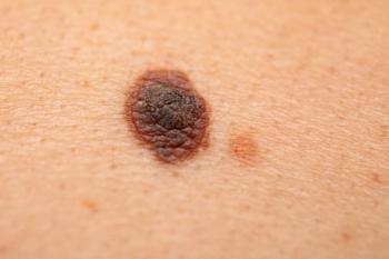
Discover how handheld Raman spectroscopy is helping distinguish skin cancer from normal tissue, courtesy of researchers from Florida Atlantic University.

FT-IR spectroscopy, long treated as a mature bench technique for confirming a carbonyl stretch or fingerprinting a polymer, has quietly become one of chemistry’s most versatile discovery engines: it now infers molecular structure straight from a spectrum without a reference library, resolves chemistry tens of nanometers wide, and screens a fingerstick of blood for disease in minutes. The last five years of published research show FT-IR moving from a confirmatory tool into a predictive, autonomous, and field-ready analytical platform.
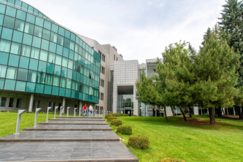
Reliability, dynamic measurements, artificial intelligence, and real-world applications are reshaping Raman spectroscopy – while Molecular Photonics points toward new translational frontiers.
Jurgen Popp, Thomas Mayerhofer, and colleagues at Leibniz IPHT and Friedrich Schiller University Jena introduce the Personalized Optical Digital Twin (PODT), a Photonics21 contribution to Europe's Virtual Human Twin ecosystem that connects molecular photonics—Raman blood analysis, coherent Raman tissue imaging, and multimodal endomicroscopy—with longitudinal physiology and clinical data. Drawing on the published multicenter INTELLIGENCE trials, the authors argue that technical feasibility and clinical utility must be evaluated separately as the field moves toward Europe's FP10 research agenda.

In this preview of the 2026 SciX Conference, we offer an overview of the keynote presentation.

A new peer-reviewed article discusses how pairing quantum mechanics with nanomaterial-based Raman spectroscopy has significantly boosted the technique's sensitivity.

Researchers developed spherical and hollow gold nanoparticle-based SERS platforms that enable label-free molecular imaging of chemotherapy drug effects and drug release within cancer cells, offering a theranostic tool with potential applications in early cancer diagnostics.
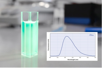
Cellular autofluorescence often masks target signals in time-resolved fluorescence imaging, and metal-based phosphors that could solve this raise toxicity and cost concerns. Thermally activated delayed fluorescence (TADF) materials offer a metal-free alternative with long emission lifetimes, and early studies show promise despite remaining challenges with quenching, solubility, and spectral purity.
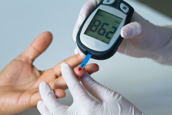
Nearly every diabetic on earth still stabs a finger to measure their blood sugar, but a small army of lasers, infrared beams, and machine-learning algorithms is quietly plotting to end that ritual for good. This review tracks the photons chasing glucose through skin, from near-infrared reflectance to Raman fingerprints, and asks why the “needle-free” promise keeps slipping just out of reach.
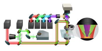
This paper demonstrates the use of optical photothermal infrared (O-PTIR) spectroscopy for the detection, identification, and localization of microplastics in tissue samples.
Advertisement
Advertisement