
Blog|Articles|January 3, 2024
SERS Used to Detect Early-Stage Liver Cancer
Author(s)Aaron Acevedo
Advertisement
In a new study published in Spectrochimica Acta Part A: Molecular and Biomolecular Spectroscopy, scientists from Kunming, China tested a new means of detecting early-stage liver cancer using surface-enhance Raman spectroscopy (SERS) (1).
Liver cancer is the sixth most common cancer and ranks third in global cancer deaths. Early liver cancer lacks obvious symptoms, and many times once a patient is diagnosed, the cancer has already progressed to middle or late stages. This has made it a priority for scientists to seek better methods for detecting the disease at earlier stages.
The two most common means of screening for liver cancer are imaging tests (ultrasound imaging, magnetic resonance imaging, computed tomography) and serological marker detection. However, these methods have certain limitations. For example, alpha-fetoprotein (AFP) is often used as a serum marker for detecting liver cancer; however, it cannot be easily distinguished in early cancer patients. Another approach, puncture biopsy, can provide a clear pathological diagnosis, though at the cost of causing patients pain.
In recent years, surface-enhance Raman spectroscopy (SERS) has become a popular approach for screening and diagnosing diseases. Notably, it is currently used to determine bioactive molecules, such as DNA, proteins, and antibodies. It offers very low detection limits and ultrahigh sensitivities at the cost of extensive preparation and selection for substrates. The comparability of SERS bases and biological samples is what most affects the technology regarding disease diagnosis. Silver (Ag) and gold (Au) are typically used as enhancement substrates; however, Ag was the preferred option for this experiment, as Ag has better absorption and conversion efficiencies that Au, while also having a better coupling effect with electromagnetic fields. But Ag is not biocompatible, meaning it can have a toxic effect on serums used in the process. This experiment was therefore conducted using Ag@SiO2 nanoparticles as SERS substrates.
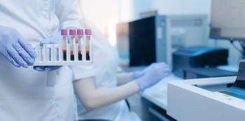
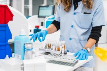
As part of the experiment, serum samples were obtained from two groups: patients with liver cancer and healthy individuals. Experiments were repeated several times, with the best SERS spectrum being obtained when the volume ratio of serum to deionized water was 1:2. From there, data preprocessing was performed on this SERS spectrum, with the data then being combined with principal component analysis (PCA), partial least-squares discriminant analysis (PLS-DA), and orthogonal partial least squares discriminant analysis (OPLS-DA) for further classification analysis. The spectral data was most effective with samples upon being combined with OPLS-DA, with a classification accuracy of 97.98%, sensitivity of 97.14%, and specificity of 98.44%. These findings highlight SERS as a potential avenue for detecting early-stage liver cancer in the future.
Reference
(1) Ou, Q.; Jinag, L.; Dou, Y.; Yang, W.; Han, M.; Ni, Q.; Tang, J.; Qian, K.; Liu, G. Application of surface-enhanced Raman spectroscopy to human serum for diagnosing liver cancer. Spectrochim. Acta A Mol. Biomol. Spectrosc. 2024, 308, 123702. DOI:
Advertisement
Related Content
Advertisement

Advertisement
Advertisement
Trending on Spectroscopy Online
1
When Political Appointees Outrank Peer Review: How a New Office of Management and Budget Rule Could Reshape Spectroscopy and Scientific Research Funding
2
The State of Spectroscopy Careers
3
From Single-Technique Analysis to Multimodal Characterization: Recent Advances and Future Perspectives for Carbon Nanotubes and Graphene
4
The Importance of Hands-On Learning
5