|Articles|June 22, 2023
Total Reflection X-ray Fluorescence (TXRF) Method Proves Effective in Analyzing Rat Tissue Samples: Preliminary Results Revealed
Author(s)Spectroscopy Staff
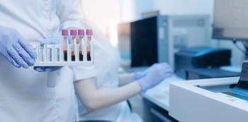
A team of researchers has conducted a successful round-robin test using total reflection X-ray fluorescence (TXRF) to analyze the elemental composition of rat tissue samples. The preliminary results demonstrate the effectiveness of TXRF in accurately determining the elemental composition of mammalian tissue.
Advertisement
Scientists from AGH University of Science and Technology in Krakow, Poland, have conducted a groundbreaking round-robin test to evaluate the element analysis of rat tissue samples using the total reflection X-ray fluorescence (TXRF) method. The study, led by Joanna Chwiej and published in Spectrochimica Acta Part B: Atomic Spectroscopy, aimed to assess the performance and reliability of TXRF by analyzing the elemental composition of rat organs in different European laboratories (1).
TXRF involves directing an X-ray beam at a shallow angle onto a sample surface coated with a thin layer of a reflective material, such as silicon. This angle causes total reflection of the X-rays at the surface, resulting in the generation of a standing wave known as an evanescent wave. As the X-rays penetrate the sample surface, they interact with the atoms of the material, causing the emission of characteristic fluorescent X-rays. The emitted X-rays are then detected and analyzed to determine the elemental composition and concentration of the sample.
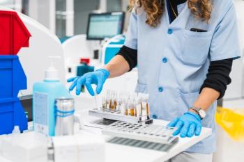
In this inter-comparison investigation, rat organs including the kidney, heart, spleen, and lung were subjected to microwave digestion. The resulting liquid samples were sent to four participating European laboratories equipped with various commercially available TXRF spectrometers. The laboratories analyzed the samples to determine their elemental composition, focusing on macroelements (P, S, and K) with concentrations above 1000 μg/g, as well as microelements (Ca) and trace elements (Fe, Cu, Zn, and Se) present at concentrations ranging from 100 to 1000 μg/g and below 100 μg/g, respectively.
The study evaluated several validation parameters, including detection limits, intra-day and inter-day precision, and inter-laboratory precision to assess the variation in results among the participating laboratories. The TXRF data was also compared with results obtained through inductively coupled plasma-mass spectrometry (ICP-MS) for Se analysis and inductively coupled plasma-optical emission spectrometry (ICP-OES) for analysis of P, S, K, Ca, Fe, Cu, and Zn.
The preliminary results demonstrated the high utility of the TXRF method for elemental analysis in animal tissues. As expected, elements with higher atomic numbers, such as Fe, Cu, Zn, and Se, yielded the best results in terms of accuracy and precision. The round-robin test confirmed good accuracy (around 100%) and precision (intra-day <6%, inter-day <12%, and inter-laboratory <12%) for these elements, highlighting the potential of TXRF for their determination in mammalian tissue samples.
However, the study also identified areas for improvement in the analysis of light elements like P, S, and K. These findings emphasized the need to optimize sample preparation procedures to minimize self-absorption in the dried residue, standardize sensitivity calibration and spectra fitting procedures among laboratories, and address other challenges to enhance the overall usefulness of TXRF in analyzing light elements.
The results of this pioneering study pave the way for future advancements in elemental analysis using TXRF and provide valuable insights for researchers working in the field of tissue analysis. By refining and standardizing analytical protocols, the TXRF method has the potential to contribute significantly to our understanding of the elemental composition of biological samples and support advancements in various scientific and medical fields.
Reference
(1) Olbrich, K.; Kubala-Kukus, A.; Marguí, E.; Fernández-Ruiz, R.; Matusiak, K.; Wudarczyk-Mocko, J.; Wrobel, P.; Setkowicz, Z.; Chwiej, J. The first total reflection X-ray fluorescence round-robin test of rat tissue samples: Preliminary results. Spectrochim. Acta Part B At. Spectrosc. 2023, 205, 106695. DOI:
Advertisement
Related Content
Advertisement


Advertisement
Advertisement
Trending on Spectroscopy Online
1
Seeing Through Stone: Spectroscopic Techniques and the Pyramids of Giza
2
Microplastics Found in All Commercial Salt Samples Tested in South Sumatra Study
3
How Are Raman, Infrared, and Mid-Infrared Spectroscopy Evolving Competitively?
4
Best of the Week: Complex Organic Molecules, Molecular Analyzers in Oil and Gas
5