SPEC PerkinElmer 11.10
|Articles|April 1, 2005

Spectroscopy

  • Spectroscopy-04-01-2005
  • Volume 20
  • Issue 4

End of the Spectrum: Science Searches for the Truth about Tut

Researchers have recently used computed tomography imaging scans to help separate fact from fiction in the ancient pharaoh's death.

Researchers have recently used computed tomography imaging scans to help separate fact from fiction in the ancient pharaoh’s death.

In hopes of solving what really killed Egypt's King Tutankhamun, better known as Tut, researchers recently performed a computed tomography (CT) imaging scan on the ancient pharaoh's remains. Now with the work complete, Zahi Hawass, Egypt's chief archaeologist and the project's leader, announced the results of the scans indicate the adolescent leader, who died at approximately 17, was not murdered as some have theorized, and that he may have died of a leg infection that came following a broken leg.

Image Courtesy of PhotoSpin

To obtain the images, a van equipped with the CT machine was parked outside of Tut's underground tomb in the Valley of the Kings in upper Egypt. Some 1700 three-dimensional images of Tut's mummy were obtained over a 15-minute span. The scans are part of the Egyptian Mummy Project, a five-year project aimed at preserving, studying, and learning from the mummies of ancient Egypt.

The imaging project comes nearly four decades after professor Ronald Harrison performed x-ray analyses on Tut's remains in 1968. Harrison pinpointed a fracture in the back of Tut's skull as the cause of his death — which grew into the theory of Tut's murder from a blow to the back of the head. Over time, this theory somehow became more fact than educated guess, and the legend of the pharaoh's murder spread.

In a recent statement released by Egyptian authorities, however, Hawass said his research team found no evidence for a blow to the back of the king's head, and no other indications of foul play. He also ruled out the theory of a possible accident that may have crushed Tut's chest. Instead, his team focused on a fracture on Tut's left thighbone as evidence of a severe injury.

"The hype about a possible homicidal death of King Tut was initiated by some loose thinking by Professor Harrison and his unsubstantiated allegations on a BBC production that the x-ray findings suggested a traumatic death," says Richard Boyer, a doctor of radiology at the Primary Children's Medical Center (Salt Lake City, Utah), who first studied Harrison's x-rays at the request of a colleague. "Unfortunately, others have bought into Harrison's fantasy and carried on the notion that the young pharaoh was murdered.

"When I was interviewed by the Discovery Channel in 2002, I spent an hour explaining the findings on the skull x-rays on camera to dispel, or at least I thought, the notion that the x-rays supported the theory of homicide," Boyer continues. "We thought that we were participating in a bona fide scientific effort to give the public some factual information on the supposed murder. We were disappointed when the Discovery Channel production aired because the producers chose to ignore all of the valid material we presented. It did not fit with their bias — it makes a lot more interesting fodder for their viewers if they can present a conspiracy theory of murder."

Now that the murder theory has been dismissed publicly by Hawass, only time will tell if the latest notion of a broken leg and an eventual fatal leg infection will hold any merit in a Da Vinci Code society that finds romanticism more appealing than scientific findings and historical facts.

"I have not seen the CT images, so I can't comment on what the leg fracture looks like. If the fracture was post-mortem, there will be no signs of healing," explains Boyer, who dismissed the erroneous murder claims in a 2003 issue of the American Journal of Neuroradiology. "The mummy was fixed into his coffin by rock-hard resin and had to be chiseled out. It would not be surprising that a leg bone might be broken in the process. After all, the neck was severed in two. If there are signs of healing of the leg fracture, then it may have occurred before death, but it would only be a source of infection if it was an open fracture, a breaking of the skin, which may be evident from a review of the scans."

Nearly 3000 years after his death, and 83 years after being discovered by British archaeologist Howard Carter, the mystery surrounding King Tut, the 12th ruler of Egypt's 18th dynasty, and his demise continues. The recent CT scans may be accepted as more dependable evidence than Harrison's x-rays, but scientists and archaeologists will have to continue their fight against hearsay and legend to have their tangible imagery and facts accepted.

"We will not reach a conclusive answer about how he died," says Boyer. "But enterprising people will keep writing and producing media materials based on shabby science as long as the public is gullible enough to buy it."

Joseph Walsh

Assistant Editor, Spectroscopy

Related to this article

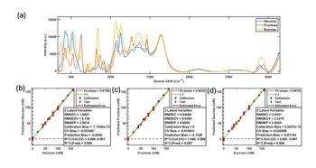
Monitoring Sugar Consumption and Ethanol Production During Yeast Fermentation Using a Raman Plate Reader and Multivariate Analysis
This column shows that a Raman plate reader paired with partial least squares regression can simultaneously quantify glucose, fructose, sucrose, and ethanol in yeast fermentation samples, using turbidity-matched calibration standards to correct for yeast-induced turbidity, and offers a rapid, label-free, high-throughput method for tracking sugar consumption and ethanol production in strain screening, media optimization, and process development.
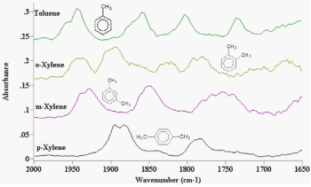
Highly Substituted Benzene Rings—The Rest of the Story II: Spectra
This column, the second installment on highly substituted benzene rings, shows that tri- and tetra-substituted substitution patterns can be distinguished by whether the ring bending peak is present and by the position of the in-phase aryl C-H wagging peak, which shifts according to how many adjacent hydrogens the ring carries.
Healthcare administrator reviewing an insurance policy schedule with a stethoscope on a clipboard, health insurance coverage, medical underwriting, prior authorization, and patient claims processing. | Image Credit: Joi_studio - stock.adobe.com
In a SciX interview, Marc Porter covered a lot of ground, from a student-driven pivot into immunoassays to a baseball analogy for Stokes and anti-Stokes scattering, and shares the open questions he is still chasing.
Ingots of pure copper or pink gold on a black background. | Image Credit: Phawat - stock.adobe.com
At SciX, Kevin Wilkinson of the Universite de Montreal explains how faster data acquisition and new software have cut analysis time and how a particle’s chemical composition can link arsenic-bearing emissions back to a copper refinery’s processes.
Spectral Frontiers: Seeing the Unseeable Microspectroscopy
Microspectroscopy, the marriage of spectroscopic chemical identification with microscopic spatial resolution, has advanced from Perkin-Elmer's first commercial infrared microspectrometer in 1953 to today's nanoscale AFM-IR probes, synchrotron-brightness infrared beamlines, and compact hyperspectral tissue imaging devices being developed for AI-assisted cancer detection.