- Spectroscopy-02-01-2020
- Volume 35
- Issue 2
New, Advanced Spectroscopic Techniques Aid in Tissue Engineering
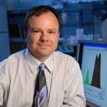
Nancy Pleshko of Temple University is studying the use of FT-IR imaging and NIR spectroscopy to solve bioengineering problems related to human replacement tissue.
Advanced techniques in tissue engineering hold promise to those who suffer from damage to or degeneration of joint cartilage. But some challenges exist for tissue engineers to gain a better understanding of the development of these constructs and their mechanical properties. Nancy Pleshko, a professor of Bioengineering at Temple University, in Philadelphia, Pennsylvania, has been studying the use of Fourier-transform–infrared imaging spectroscopy (FT-IRIS) as well as near-infrared (NIR) spectroscopy to explore the ways in which these techniques can aid in the development of replacement tissue. We spoke to her about her research and findings.
In a recent paper about tissue engineering and its ability to repair defects in articular cartilage (1) you discussed the use of FT-IRIS to study the distribution of water and extracellular matrix produced in engineered constructs such as those that are created for cartilage repair. Why is this an important study, and how might it help patients with osteoarthritis?
Tissue engineering strategies are being investigated for repair of tissues that are challenging to regenerate naturally, such as articular cartilage. Osteoarthritis is a disease that results in degeneration of joint (articular) cartilage, resulting in pain and mobility loss for millions. Development of engineered constructs could be a solution for tissue repair, but the compositional properties of the native cartilage should ideally be duplicated in the engineered tissues. The primary tissue components are extracellular matrix, including collagen and proteoglycans, and water. Thus, we need to know the relative amount and distribution of these components in both native tissue and engineered constructs.
What are the advantages of using FT-IRIS over more traditional techniques that allow a visual comparison between native cartilage and engineered cartilage? You reported spatial resolution of 25 mm to permit visualization of matrix and water throughout the tissue. What would be the ideal spatial resolution for this work?
There are several advantages of using this technique. First, FT–IRIS permits quantification of compositional information without the addition of external contrast agents. Different protein components, or mineral, if present, and water, can be evaluated in one unstained tissue section. This saves a significant amount of tissue preparation time and preserves the rest of the tissue for additional studies. Further, many other techniques can’t be used to visualize water distribution, so this is certainly an advantage of this modality. The ideal spatial resolution would vary, depending on the question of interest. In cartilage, for example, spatial resolution in the 10–25 mm range is acceptable, as this can be related to variations in different zones of cartilage. However, it could be desirable to have better spatial resolution if there is a question of what specifically a cell is producing. Then one may want to investigate this at a better resolution, which could theoretically be done with attenuated total reflection (ATR) spectral imaging.
What challenges did you face in developing this method? Did you encounter problems with sample preparation and cryosectioning 80-mm sections of native and engineered cartilage?
The primary challenge was optimizing the sample thickness to enable collection of data from the same sample in both the mid-IR and NIR spectral regions. The sample had to be thick enough to obtain a reasonable NIR signal, but not so thick that the mid-IR absorbances were off scale. Investigation of mid-IR absorbances related to collagen and proteoglycan that have small absorptivity coefficients enabled us to achieve this goal. But, protein absorbances that are typically investigated in the mid-IR, such as the amide I absorbance, would not be feasible in an 80-mm tissue section.
Another recent paper of yours describes how NIR spectroscopy may be an alternative to destructive biochemical and mechanical methods for the evaluation of hyaluronic acid (HA) (2). What makes NIR a good technique for evaluating HA?
In that study, we used an NIR fiber optic to investigate hyaluronic acid–based developing engineered cartilage tissues. Typically, engineered tissues have to be harvested for implantation at an optimal timepoint, and determination of that timepoint is challenging. In particular, if, for example, you’re growing six constructs, you may harvest one, and perform a biochemical analysis to assess the composition, and whether or not the construct is ready to implant. However, the variation in composition among constructs can be high, even if grown in the exact same conditions. So, relying on the outcome from one construct to reflect the composition of another in the same batch isn’t always accurate. Thus, collection of NIR fiber optic data that reflect compositional data of an individual construct is an ideal solution.
What results have you achieved so far, and what differences, if any, can you observe between native and engineered cartilage using FT-IR imaging and NIR?
So far, we have successfully developed multivariate models based on NIR fiber optic data to predict compositional properties of developing engineered cartilage. We have also used FT-IR imaging data to help us understand how the fiber optic data change based on distribution of various components in the tissues, and how sensitive the fiber optic data are to these variations. The combination of the two different sampling modalities enables us to more fully understand the tissue development, and how to optimize our data collection and processing.
What are your next steps in this work?
The next steps for us are going to be very exciting! We will grow engineered cartilage in the laboratory, identify optimal constructs based on our NIR fiber optic data, and then harvest them to implant in a preclinical model. Further, we will use the same fiber optic sampling to assess how the engineered construct composition changes once it is implanted in vivo. If these experiments are successful, it will be a major step towards improved ability to select ideal constructs for tissue repair.
References
- J.P. Karchner, W. Querido, S. Kandel, and N. Pleshko, Ann. NY Acad. Sci.442(1), 104–117 (2019). doi:10.1111/nyas.13934
- F. Yousefi, M. Kim, S.Y. Nahri, R. L. Mauck, and N. Pleshko, Tissue Eng. Regen. Med.24(1,2) (2018). doi:10.1089/ten.tea.2017.0035
Articles in this issue
about 6 years ago
Vol 35 No 2 Spectroscopy February 2020 Regular Issue PDFabout 6 years ago
Use of Raman Microscopy to Characterize Extractables and Leachablesabout 6 years ago
Raman Spectroscopy of Graphene-Based Materialsabout 6 years ago
LIBS-Based Imaging: Recent Advances and Future Directionsabout 6 years ago
Market Profile: Near Infrared (NIR) SpectroscopyNewsletter
Get essential updates on the latest spectroscopy technologies, regulatory standards, and best practices—subscribe today to Spectroscopy.


