Surface-Enhanced Raman Spectroscopy for Improved Disease Detection
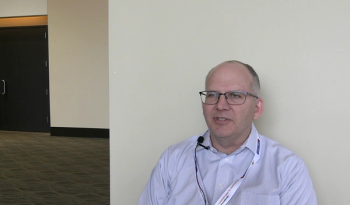
Surface-enhanced Raman spectroscopy (SERS) is an exciting avenue of study in the field of disease research, particularly with respect to its potential ability to provide enhanced detection compared with previous analytical techniques. Marc D. Porter, who is a professor of Chemistry and Chemical Engineering at the University of Utah, has been working with SERS to improve the detection of diseases such as tuberculosis and hepatic cancer. We recently spoke with him about this research.
Surface-enhanced Raman spectroscopy (SERS) is an exciting avenue of study in the field of disease research, particularly with respect to its potential ability to provide enhanced detection compared with previous analytical techniques. Marc D. Porter, who is a professor of Chemistry and Chemical Engineering at the University of Utah, has been working with SERS to improve the detection of diseases such as tuberculosis and hepatic cancer. We recently spoke with him about this research.
One of your recent research projects involved the use of SERS to study a lipoglycan component of the cell wall of Mycobacterium tuberculosis, the pathogenic bacterium responsible for tuberculosis (1,2). Specifically, SERS was used to monitor the effects of sample pretreatments intended to counter the complexation of the lipoglycan with antibodies and other proteins in serum (complexation decreases the limit of detection for the lipoglycan). Can you please briefly describe the SERS approach used in this study?
The approach adopts a construct much like that of a sandwich enzyme-linked immunosorbent assay (ELISA). In our case, we use a capture substrate that consists of a thin film of smooth gold (~200 nm) coated onto a silicon wafer, which is addressed with an adsorbed layer of antibodies specific for the lipoglycan. Like ELISA, in the first step, we pipette a small volume of a serum sample (for example, 20 µL) onto the capture address, and the layer of antibodies then selectively binds the antigen from the sample. We then rinse the surface and treat it with a suspension of what we call extrinsic Raman labels or ERLs. ERLs are gold nanoparticles designed to selectively tag captured antigen and to generate a strong Raman signal upon plasmonic excitation with laser light at 633 nm. These two functions are imparted by first coating the gold nanoparticles with monolayer of a Raman reporter molecule, or RRM, and then an adsorbed layer of tracer antibodies. The binding specificity of the tracer antibodies recognizes the captured antigen, which is indirectly signaled by the spectrum of the RRM. We use the strength of the strongest spectral feature of the RRM for antigen quantification. The difference is that the signal we measure is amplified plasmonically, whereas response for ELISA is generated by catalytic generation of a colored product by the action of an enzyme conjugated to the tracer antibody.
What makes SERS a good choice for assessing the sample pretreatment effects?
There are actually two parts to the answer to this question. The first part reflects the approach we use in making a preliminary assessment of how well our SERS platform may work when attempting to detect a new marker. This work begins by spiking a buffer like phosphate-buffered saline (PBS) with the marker and running an assay to get preliminary data for limit of detection and analytical sensitivity. If the results look promising with respect to the literature, we then design a study to determine how well the platform works in serum or urine. The second part of the answer is that we can typically detect a marker with antibodies of reasonable binding affinities at low picomolar levels or lower, and it is normal for us to lose a bit of detectability when moving from buffer to serum. So, when we began the work on the lipoglycan in our tuberculosis project, we were surprised by the fact that detectability degraded by a factor of 1500×. This loss in detection served as a clue that something was amiss when attempting to measure this marker in serum; and, let me add that attempts to measure this marker in urine reveled that there was an unrecognized detection problem there too.
Your research showed that a simple pretreatment process improved the limit of detection for the lipoglycan in serum by 250-fold, which demonstrates the potential of the lipoglycan’s use as an antigenic marker for tuberculosis (2). What are the advantages of the SERS approach compared with other methods for detecting antigenic markers of diseases such as tuberculosis and malaria?
There are several drivers here. First, as I mentioned earlier, we can typically detect markers with SERS at picomolar levels, and often down to femtomolar levels. Our past work, as well as those of others, have in fact, shown that SERS often rivals or surpasses that of fluorescence, in some instances. What motivates us in our work is that we can realize this capability without the need of the incubation step in ELISA. Second, we favor the use of gold as our plasmonic material, allowing us to excite the signal with a long wavelength source (e.g., 633 nm or 785 nm). This approach often eliminates any native fluorescence from the sample matrix, which can be a major interference with SERS when using short wavelength excitation materials. Third, the line widths of vibrational spectra are 10–100× narrower than those encountered when using an assay with a readout based on excited electronic state spectroscopies. Raman levels of spectral overlap are markedly smaller. This characteristic enables the ability to multiplex the detection of more markers when using spectrally distinct RRMs, if you can avoid problems with antibody cross reactivity. Fourth, our SERS assay requires smaller amounts of sample, reagents, and other materials when compared to ELISA and several other popular forms of immunoassays.
What is the potential impact of your approach on the field of tuberculosis research?
We believe that by taking advantage of the detection capabilities of our SERS platform when combined with the impact of sample preparation, the point-of-need (PON) embodiment that we have been recently working on has the potential to serve as the underpinnings of a portable and low-cost test that be used in the global fight against this horrible disease.
A second study involved the development of a portable immunoassay method that combines solid-phase extraction membranes, gold nanoparticle labels, and SERS for hepatic cancer screening (3). Can you please outline this approach?
This work was aimed specifically at looking for a way to move our SERS platform from the research laboratory to PON. It is actually a redesign of colorimetric solid-phase extraction, or C-SPE, technology that was deployed for potable water analysis on the International Space Station (ISS) several years ago. C-SPE is designed to rapidly and selectively concentrate an analyte in a manner like the high capacity, microporous membranes and particulate phases used so successfully by solid-phase microextraction (SPME). Most of the time, SPME is used in measurements by mass spectrometry or other types of analysis. It is largely the high concentration factors of SPME, up to 1000 or more, that resulted in its use in a number of different types of analyses. With C-SPE, the analyte can be quantified on the membrane after its selective complexation and concentration with a colorimetric reagent impregnated in the membrane.
In adapting C-SPE to work with SERS, we simply impregnate a capture reagent in the microporous membrane. This work also took a different approach to how the liquid sample is passed through the membrane. Instead of using a handheld syringe, an adsorbent paper is affixed to the bottom of the membrane to pull the liquid sample through the membrane by capillary action. The antigen-tagging step simply repeats the process, but with a small volume of ERL suspension. This approach allows us to combine the attractive sampling features of SPME with the ultrasensitive detection strength of SERS.
What is the ultimate goal of this study?
This part of our work is focused on the development of a screening panel to identify individuals at risk of developing hepatocellular carcinoma (HCC). HCC is the third highest cause of cancer-related mortality worldwide. In an all too familiar theme, this high mortality rate is due to its diagnosis at a stage of progression beyond which resection and other potential curative interventions are effective. However, research indicates that early detection followed by surgical resection can dramatically improve survival rates. Our goal is to develop a screening panel that has a multiplexed marker signature that can be used a means for the reliable at-risk patient assessments. Our strategy calls for the development of a multiplexed screening panel of five important serum markers identified in liver cancer patients. Our work in the first stages of this project have shown that by integrating SERS with SPME, we can measure one critical marker, alpha fetoprotein (AFP), from a small volume (10 mL) of human serum at a limit of detection of roughly 40 fM. While this level of detection surpasses that considered today as the cutoff between healthy and at-risk individuals by 1000×, these results are beginning to demonstrate the strengths of this technology as a potential addition to the point-of-need toolbox.
Your group recently looked at the use of a handheld Raman spectrometer for quantitative tuberculosis biomarker detection (4). That study compared results obtained using a field-portable spectrometer with those obtained using a much more sensitive benchtop Raman spectrometer. How did the limits of detection for a nonpathogenic antigenic marker simulant compare using the two systems?
The results showed that the detection capabilities of a handheld spectrometer are approaching those needed to aid treatment decisions in tests carried out in remote locations for tuberculosis and possibly other infectious diseases. For this, we compared measurements of a field-portable Raman spectrometer with those obtained using a much more sensitive benchtop Raman spectrometer for a TB biomarker simulant. While not an exact duplication of measurement conditions, our findings were promising. We found that the limit of detection with the handheld system (~0.2 ng/mL) approached that of the benchtop instrument (0.03 ng/mL). More so, this work indicated that if the conditions for plasmonic amplification were tuned to match those of the excitation wavelength of the handheld spectrometer, the two types of spectrometric systems become much more comparable in terms of detection strength.
What challenges remain before your SERS immunoassay can be implemented in point-of-need applications?
This is another answer that has two parts. The first is about the clinical accuracy of the assay itself. Can we, in fact, design SERS assays for different markers at a high enough level of clinical accuracy and reliability to gain the trust and confidence of a caregiver for them to incorporate the data from an assay into their treatment decision making? I, along with many of my colleagues in the area of diagnostic test development, believe the answer is an unquestionable yes. But, and this is the a big but, we need to stay the course and invest the needed sweat equity to chase down the myriad challenges yet unsolved in fully realizing the reliability of SERS as an invaluable analytical measurement tool. We must also enlist the help of our colleagues in the clinical diagnostics arena to develop procedures that fit in the existent workflow in their laboratories and to run validation tests on sufficiently large numbers of patient samples to demonstrate the clinical value of this technology over what is in use today.
What are the next steps in your research?
In addition to working on how to move our platform closer to PON, there are a number of new avenues to explore, both fundamentally and technologically. At the fundamental level, we are still delinquent in putting together a theoretical model of the assay that incorporates the kinetics of antigen binding and ERL tagging, and it is still unclear to me how to truly think about the ERL tagging process. Can we, for example, view tagging a monovalent (1:1 reaction stoichiometry) process, or is there a level of multivalency that is important but not yet recognized? We have also just learned that gravity plays a much more important role in the accumulation of ERLs on the capture substrate than we first thought, and that the impact of ERL sedimentation can add to the level of the background response and, therefore, degrade detectability. What else have we missed or overlooked?
The next immediate technological hurdles for us center on sample preparation. We think that immunocomplexation and other processes play a much larger role in these tests than previously recognized in compromising the clinical utility of a number of disease markers, and we are working on ways to begin to test this perspective.
I would like to thank you for the opportunity to talk with you about our work in this area. It has been both interesting and thought-provoking!
I would also like to acknowledge the people I have had the good fortune of working with over the course of these projects and those who before set the stage for our recent work. While I understand that I will not be able to personally recognize all of them, I would like to give them all a shout out: Thank you!
References
1. L.B. Laurentius, A.C. Crawford, T.S. Mulvihill, J.H. Granger, R. Robinson, J.S. Spencer, D. Chatterjee, K.E. Hanson, and M.D. Porter, Analyst (2016). doi: 10.1039/c6an02109c
2. A.C. Crawford, L.B. Laurentius, T.S. Mulvihill, J.H. Granger, J.S. Spencer, D. Chatterjee, K.E. Hanson, and M.D. Porter, Analyst (2016). doi: 10.1039/c6an02110g
3. J.H. Granger, A. Skuratovsky, M.D. Porter, C.L. Scaife, J.E. Shea, Q. Li, and S. Wang, Analytical Methods9, 4641 (2017). doi: 10.1039/c7ay01689a
4. N.A. Owens, L.B. Laurentius, M.D. Porter, Q. Li, S. Wang, and D. Chatterjee, Applied Spectroscopy (2018). doi: 10.1177/000370281877066
Newsletter
Get essential updates on the latest spectroscopy technologies, regulatory standards, and best practices—subscribe today to Spectroscopy.


