
News|Articles|January 5, 2024
Using SERS to Explore Cancer Cells with MTAP Deletions
Author(s)Spectroscopy Staff
A recent study from Spain used surface-enhanced Raman spectroscopy (SERS) to study cancer cells with methylthioadenosine phosphorylase (MTAP) deletions, shedding new insights into the metabolic interactions inside the tumor microenvironment that could influence cancer aggression.
Advertisement
Oncology, which is the study of cancer, is one of the largest healthcare disciplines. Understanding how cancer develops in the human body is paramount to improve human health and save lives. One of the main topics in oncology that researchers are exploring is cancer aggression, or how molecular changes in cancer cells and the tumor microenvironment (TME) can affect the aggressiveness of how cancer spreads in the human body.
A recent study published in PNAS explores this very question. A team of researchers led by Arkaitz Carracedo and Luis M. Liz-Marzan from the Center for Cooperative Research in Biomaterials (CIC biomaGUNE) examined cancer cells with methylthioadenosine phosphorylase (MTAP) deletions using surface-enhanced Raman spectroscopy (SERS) to further understand the metabolic interactions that take place inside cancerous cells and the TME (1).
The TME is an important organ in cancer research because it often dictates how fast or slow the cancer spreads in the body. Using SERS, the research team discovered that cancer cells with MTAP deletions exude 5′-methylthioadenosine (MTA) (1). The production of MTA, coupled with fibroblasts that metabolize it, results in molecular changes that directly influence the progression of cancer, affecting the polarization of macrophage (1).
Several changes occur when MTA interacts with fibroblasts, which the authors describe in their study. First, the capacity of purine derivatives to induce macrophage polarization is impacted (1). Second, the molecular changes that occur accelerate the aggression of cancer, resulting in it spreading more quickly (1). Finally, the production cycle of purine derivatives is changed and can be recycled by cancerous cells (1).
Previous studies in this work focused on protein messengers. Traditional methods used for profiling metabolites in cells are limited (1). The use of SERS in deciphering metabolic interactions in multicellular systems has not been explored much, but because of the technique’s simple operation and nontargeted nature, it is optimal for application in metabolic research (1).
In this study, the researchers demonstrated in this study that SERS could be applied when analyzing secreted metabolites in the TME (1). By using SERS in this context, the hope is that it could innovate existing therapeutic strategies to improve them for future generations.
This article was written with the help of artificial intelligence and has been edited to ensure accuracy and clarity. You can read more about our
Reference
(1) Valera, P. S.; Plou, J.; Garcia, I.; et al. SERS analysis of cancer cell-secreted purines reveals a unique paracrine crosstalk in MTAP-deficient tumors. PNAS 2023, 120 (52), e2311674120. DOI:
Advertisement
Related Content
Advertisement


Advertisement
Advertisement
Trending on Spectroscopy Online
1
When Political Appointees Outrank Peer Review: How a New Office of Management and Budget Rule Could Reshape Spectroscopy and Scientific Research Funding
2
UV–FT-IR Studies on α-Lactalbumin-Infused Nickel Hydroxide Nanoparticles
3
What is the Gordon Research Conference on Vibrational Spectroscopy?
4
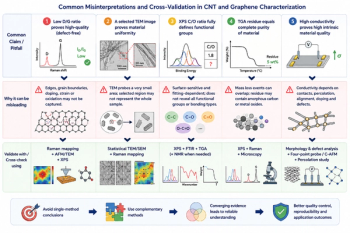
From Single-Technique Analysis to Multimodal Characterization: Recent Advances and Future Perspectives for Carbon Nanotubes and Graphene
5