Article
Special Issues
Spectroscopy Supplements
Raman Spectroscopy of Documents
Author(s):
Documents have been investigated to determine the feasibility of utilizing Raman and SERS Raman spectroscopy for the identification and characterization of inks on paper. Fluorescence reduction methods have been employed to facilitate the analysis by reducing the nascent fluorescence from paper and ink. Furthermore, ink crossings were investigated to demonstrate that ink applied after creation of a document could be differentiated from the originally applied ink.
Raman spectroscopy has been shown to be important in the forensic analysis of trace evidence, controlled substances, and documents (1). Document analysis can be broken down into a few categories of investigation, including authentication of documents of value, determination of document alteration, and association of evidence to a suspect or place of origin. Because Raman spectroscopy, like infrared spectroscopy, accesses fundamental modes of vibration, it is highly specific for chemical identification. The spectral resolution of Raman spectroscopy is typically higher than that of condensed phase infrared spectroscopy, further aiding in the discrimination of molecular species. It is worth noting that Raman analysis is nondestructive and allows in-situ analysis even when applied to investigations in aqueous solutions. Infrared microscopy measurements are typically limited at longer wavelengths, due to the use of photoconductive mercury-cadmium-telluride (MCT) detectors that have a long wavelength cutoff of about 600 cm-1 . Modern Raman microscopes routinely allow the detection and identification of inorganic pigments that frequently have spectral signatures down to ~100 cm-1 .

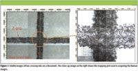

Figure 1
There are a few obstacles to the successful Raman analysis of documents. First, paper frequently exhibits fluorescence upon visible excitation, where the fluorescence signal dwarfs the Raman signal. Secondly, many synthetic dyes found in textiles, inks, and paints have inks and pigments that also exhibit fluorescence. Third, many inks are dark in color and subsequently undergo transient heating upon impingement of the incident laser light. This can yield damage or alteration of the document in question. Fluorescence reduction or rejection methods must therefore be frequently employed to collect useful Raman spectra. The first method employed for this investigation is the use of a concave baseline correction method that eliminates the perturbing fluorescence spectrum that is superimposed on the desired Raman spectrum (2). The second method used was surface-enhanced Raman scattering (SERS). The SERS effect is characterized by an increase in the Raman intensity by many orders of magnitude for species adsorbed on rough metal (usually silver) surfaces compared to that obtained from the same number of molecules in solution or the gas phase (3,4). At the same time, proximity to the surface provides a nonradiative pathway for relaxation from the excited state, which successfully quenches fluorescence. Recent work in SERS has demonstrated that organic colorants present in inks, paints, and textile fibers can be easily identified from microscopic samples by treatment of the sample with silver nanoparticles before analysis (5). This method has proven effective for the detection of specific dyes from samples as small as a 1-mm section of a single silk fiber of 50-μm diameter and on dyes used in textiles (on archaeological samples) even when severely degraded by burial. Emerging methods for completely nondestructive SERS analysis of documents and textile fibers will enable the identification of inks and dyes while preserving the integrity of evidence such as textile fibers or documents.


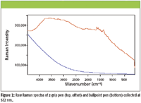
Figure 2
Application Examples
One area of interest is the detection and characterization of ink applied after the creation of a document. This would be important in determining whether a document has been altered and, if so, which inks were applied first. Confocal Raman microscopy can be applied such that a depth profile can be performed on the ink in question to determine which occurred first. The depth resolution for confocal Raman microscopy is about 1 μm. If ink is applied first and allowed to dry, subsequent application of ink would sit on top of the earlier deposited ink. If an ink applied later is composed of a different pigment, Raman would readily differentiate between the inks. Figure 1 shows an ink crossing for two different inks applied within a few seconds of each other. Figure 2 shows the raw Raman spectra collected from a ballpoint pen (top) and a z-grip pen (bottom). The Raman spectra were collected using a Senterra Raman microscope (Bruker Optics, Inc.) at 532 nm with a 1-s integration in the confocal mode of operation at a resolution of 3 cm-1. The microscope was mounted on a z-stage to allow access to large samples. Those same Raman spectra are shown in Figure 3 after removal of the fluorescence by utilization of the concave baseline correction method. Before fluorescence removal, the overlaying Raman spectra are barely observable. The corresponding Raman images are shown in Figure 4, where the 1648 cm-1 band was integrated for the z-grip image and the 1146 cm-1 band for the ballpoint pen image. The ballpoint pen was readily identified as methyl violet and the z-grip ink as methyl violet with an additive. It is clearly evident that the z-grip pen was applied over the ballpoint pen.

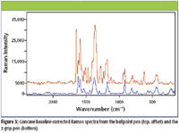

Figure 3

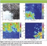

Figure 4
We can extend the investigation further by bringing the document through the field of focus to depth resolve each of the inks at the junction point, as shown for the z-grip pen in Figure 5. The depth profile was conducted by stepping in 1-μm steps from the top surface focus position into the sample for a total of 100 μm. As expected the spectrum at the interface was composed of a combination of the spectra representative of both inks. This example demonstrates the facility of confocal Raman microscopy for document analysis, so long as the fluorescence can be effectively removed.

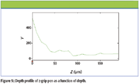

Figure 5
Another area of forensic interest is the analysis of documents generated from inkjet printers. Figure 6 shows the visible images from such a document. The resultant Raman spectra also exhibited significant fluorescence, where concave baseline correction was also employed. The Raman images, shown in Figure 7, demonstrate the ability of Raman spectroscopy to be used for the characterization of documents via pigment identification.

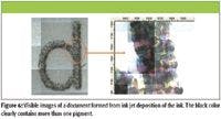

Figure 6
Another document was examined with SERS to determine if the fluorescence could be quenched effectively as well as achieve a signal enhancement. This document had ink containing a phthtalo blue and a methyl violet ink. The 488-nm spectrum shows some peaks of both over a fluorescent background. The quality is low due to the paper fluorescence, and to the methyl violet fluorescence. Going to 785 nm does not improve things much because, as the fluorescence of methyl violet decreases, phthalo blue becomes slightly fluorescent. In this case, a droplet of silver colloid suspension, prepared using a previously described method in which microwave-assisted reduction is employed, was deposited onto the document using a piezo device prior to analysis. The silver colloid droplet size was about 20 μm in diameter. 488 nm excitation was used for the analysis of this document. Figure 9 shows the Raman spectrum without correction (top) and the SERS result (bottom). It is clear that the fluorescence has been quenched effectively and a very high quality SERS Raman obtained. This striking example shows the promise of using SERS Raman microscopy in the detection of very small amounts of dyes and pigments even on fluorescing substrates such as paper or skin.


Figure 7
In summary, Raman microanalysis can become an important tool in the forensic analysis of documents. Previously, the interference from fluorescence precluded the widespread application of Raman to this field of study. With the advent of current state-of-the-art methods for removing fluorescence and the emergence of SERS Raman as a viable and practical method for quenching fluorescence and enhancing the Raman signal, it is expected that interest and activity in this field will increase greatly.
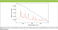

Figure 8
Thomas J. Tague Jr. and Peng Wang are with Bruker Optics, Inc., Billerica, Massachusetts. Marco Leona is Head of Scientific Research with the Metropolitan Museum of Art, New York, New York.
References
(1) E.G. Bartick, Handbook of Vibrational Spectroscopy (2002).]
(2) M. Pirzer and J. Sawatzki, US Patent 2006/0212275.
(3) J.R. Lombardi, R.L. Birke, T. Lu, and J. Xu, J. Chem. Phys. 84, 4174 (1986).
(4) C. Rodger, G. Dent, J. Watkinson, and W.E. Smith, Appl. Spectrosc. 54(11), 1567–1576 (2000).
(5) N. N. Daeid (Editor), Review Papers, 14th International Forensic Science Symposium (2004).
(6) M. Leona, J. Stenger, and E. Ferloni, ArtRaman 2005 3rd International Conference on the Application of Raman Spectroscopy in Art and Archaeology (2005).
(7) M. Leona, Proc. Nat. Acad. Sci. USA106, 14757–14762, 2009.(8) M. Leona and T.J. Tague Jr., US PatenPending.


Newsletter
Get essential updates on the latest spectroscopy technologies, regulatory standards, and best practices—subscribe today to Spectroscopy.