Advancing Materials Analysis with Imaging
For the characterization of complex materials, imaging methods such as atomic force microscopy, scanning Kelvin probe microscopy, luminescence-based hyperspectral imaging, and confocal Raman microscopy-sometimes in combination-are used to obtain sufficient details about chemical heterogeneity across multiple length scales.
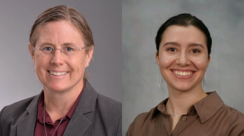
Frank V. Bright, the Henry M. Woodburn Chair and a SUNY distinguished professor of chemistry at the University of Buffalo, the State University of New York, has been using these imaging techniques in his research, for which he won the 2015 ACS Award in Spectrochemical Analysis. He recently spoke to us about his work applying these techniques to the analysis of antifouling films-designed to prevent algae and other marine organisms from accumulating on ships-and to the forensic analysis of fibers.
In a recent paper (1), you described using colocalized atomic force microscopy (AFM), scanning Kelvin probe microscopy, and confocal Raman microscopy to analyze a model antifouling xerogel film designed to prevent the spread of invasive species in a marine environment. What spectroscopic techniques have been used in the past to analyze antifouling films, and what advantages did your approach provide over past approaches for topographic and chemical analysis of the film?
In previous research we had used AFM and IR imaging. However, those experiments were not carried out in a colocalized manner so what we observed in AFM was not the exact same position observed by IR. Further, although the AFM offered nanometer spatial resolution or better, the IR measurements allowed a spatial resolution of 3–4 µm. Thus, features that were smaller than 3–4 µm could not be resolved or assessed fully.
By using colocalized AFM, scanning Kelvin, and Raman we are studying the same physical positions on the sample in a colocalized manner and measuring topography, surface charge, and vibrational spectroscopy information simultaneously, and the overall spatial resolution is on the order of 250–300 nm-the Raman diffraction limit. Using these techniques together allowed us to correlate topographic and surface charge with specific chemical species with 250–300 nm resolution.
What challenges did you face in this study?
The experiments per se were straightforward. One challenge arose when we observed a feature in Raman that was not seen at all in AFM or scanning Kelvin. Raman revealed that this feature was associated with a hydrogen-bonded amine. After some thought, we realized that this feature was buried just below the xerogel film surface and we saw it by Raman because the voxel depth allowed us to look into the xerogel material to a depth of up to 600 nm in our case. Thus, the AFM and scanning Kelvin experiments see only the surface whereas the Raman sees the surface and things buried just below the surface.
In another recent study (2), you used luminescence-based hyperspectral imaging to study denim fiber bundles. You and your group used principal component analysis (PCA), red-green-blue histogram analysis, and a one-way analysis of variance (ANOVA) to examine the data. Which of these data analysis techniques proved to be the best at discriminating between the fiber bundle types, and why?
It turned out that PCA was the best of the methods tested. I think the reason is associated with PCA using the entire spectrum whereas the red-green-blue analysis parsed the spectrum into just three primary colors.
Do you expect the hyperspectral imaging technique to eventually be adopted by forensic chemists for fiber-bundle analysis?
Perhaps. I can see it being used on individual fibers with higher magnification. I think the part of the paper that might be implemented first is the use of CH3NO2 quenching. We showed that CH3NO2 partitions into the fiber materials, quenches the fluorescence, and does so in a reversible manner. The key is that the CH3NO2 does not partition into the bundle uniformly. Thus, one gains additional contrast when using CH3NO2.
What are your next steps in your research?
Our research program is focusing on developing chemical sensors based on silicon nanostructures (silicon quantum dots and porous silicon), studying antifouling films that function because they possess chemically and topographically ambiguous domains, and understanding the interaction of exogenous agents with the corneal epithelial surface. In all these areas, imaging methods are playing an ever-increasing role, and we are moving more and more into the imaging, be it using optical methods like those I discussed earlier, or imaging based on X-ray photoelectron spectroscopy or secondary ion mass spectrometry.
(1) J.F. Destino, C.M. Gatley, A.K. Craft, M.R. Detty, and F.V. Bright, Langmuir 31, 3510–3517 (2015).
(2) R.E. Deuro, K.M. Leiker, Y. Wang, N.J. Deuro, T.M. Milillo, and F.V. Bright, Applied Spectroscopy69(1), 103–114 (2015).
Newsletter
Get essential updates on the latest spectroscopy technologies, regulatory standards, and best practices—subscribe today to Spectroscopy.


