Artificial Intelligence’s Potential in Microscopy: An Interview with Ana Doblas of the University of Massachusetts Dartmouth
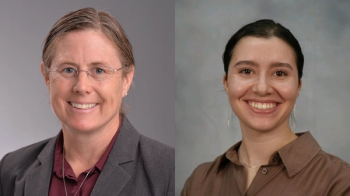
At SPIE Photonics West, Spectroscopy spoke with Ana Doblas of the University of Massachusetts, Dartmouth about artificial intelligence, deep learning, and their potential in microscopy.
Artificial intelligence (AI) and machine learning (ML) have emerged as useful tools for spectroscopists who want to analyze large data sets relatively quickly. Spectroscopy recently caught up with Ana Doblas, the principal investigator of the Optical Imaging Research Laboratory at the University of Massachusetts, Dartmouth, at the SPIE Photonics West conference to discuss how AI and playing a bigger role in microscopy.
Doblas's current research interests are focused on optical engineering, computational optics, and three-dimensional imaging with a special interest in the design of novel microscopic imaging systems and their applications (1). The mission of her lab is to integrate research and education to stimulate interest in optical engineering, providing students with a unique set of skills for designing and building future technologies in Optics and Photonics. Since 2012, she has been the author of 40 peer-reviewed scientific journal articles, her work has been presented at more than 80 international conferences, and she is co-inventor of 3 US patents.
In this interview, Doblas discusses deep learning, specifically digital lensless holographic microscopy (DLHM), and its uses in reconstruction algorithms, as well as how artificial intelligence can be used in microscopy imaging and microscopy in general.
Q: What is DLHM? Can you explain how it works?
A: DLHM is a really a fancy term to basically describe the light propagation or diffraction. In other words, a digital lensless holographic microscope is basically a spherical waveform illuminating a sample, and then we record the diffraction pattern or what some people call the lensless inline holographic image. The recorded image is not an in-focus replica of the sample. But, if the sample is placed close enough to the sensor/camera, then the recorded image is quite like the in-focus one.
Q: Why is using DLHM better than using other types of lenses/hardware?
A: I would define “better” more as “simple, compact, and cheap” since one only needs a camera and a source to illuminate your sample. So, it’s super simple and super convenient, though its resolution capability cannot be compared with the microscopic systems that use a microscope objective lens with a high numerical aperture. Nonetheless, some applications do not require such resolution, making the DLHM technique a suitable one for them.
Q: What are examples of deep learning models replacing traditional DLHM reconstruction algorithms?
A: After one records the defocused image in a DLHM system, a computational algorithm is required to perform the back-propagation of the light to reconstruct the in-focus sample distribution. My colleagues, Carlos Trujillo, PhD, from the University of EAFIT in Colombia and Jorge Garcia-Sucerquia, PhD, from Universidad Nacional Sede Medellin in Colombia, have recently worked in a deep learning model in which a convolutional neural network (CNN) based on regression model predicts the axial position of the sample from the raw defocused image, reducing the computational burden of the search of the in-focus image and enabling fast reconstruction of DLHM images. Other examples are made by Aydogan Ozcan’s group from UCLA. Typically, one should reconstruct the in-focus images before classifying the sample. Jointly with Trujillo, we are currently working on the classification of sample types from the raw images, without any reconstruction algorithm. So, the question that we are posing right now is, do the diffraction patterns that we are recording have enough classification power so that we can classify the sample without reconstruction them?
Q: What applications does AI have in microscopy imaging?
A: There are different kinds of applications. One of them is using AI as a computational tool for counting cells or segmenting images. Other example is the identification of cancer cells. For example, cancer cells are identified by a trained physician by analyzing the presence of an emitted fluorescent signal in the recorded image using typically a confocal scanning microscope. This process can be performed faster if the data analysis is provided by an AI model instead of a person. Over the last five years, AI has been proposed as a reconstruction tool, replacing traditional image processing frameworks. The quality of the reconstructed images in traditional image processing frameworks is highly dependent on the person reconstructing the image. For example, quantitative phase images require some intuition to determine if the reconstructed phase images present phase distortions or not, requiring trained people to achieve the best quality of image. I believe that AI models can provide high-quality images at a faster time, independently on the knowledge of the user. This is a key part if we want to transfer our methods to biological researchers, who do not have any knowledge about imaging processing algorithms.
Q: What benefits do you see of AI in the microscopic field?
A: Connecting to the end of the last question, AI can help us to reconstruct high-quality images independently on the experiences of graduate students. It could also help us to improve the collaboration between imaging researchers and biological researchers, providing user-friendly techniques. Another potential benefit is the reduction of the price. If we can get the same performance of a cheap system boosted with AI is the same as its analogous commercial high-performance system that costs more than $100,000, I think that we could bring these high-performance imaging modalities to more users. This is a significant benefit of AI in the current microscopy field.
Reference
(1) UMass Dartmouth. Ana Doblas, PhD. Assistant Professor at the Department of Electrical and Computer Engineering, University of Massachusetts Dartmouth.
Newsletter
Get essential updates on the latest spectroscopy technologies, regulatory standards, and best practices—subscribe today to Spectroscopy.


