How did the researchers test their technique?
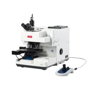
The research team tested their technique by using C57BL/6 mice as animal models, simulating death by strangulation and drowning. Then, the lung tissues were collected and analyzed postmortem using ATR-FTIR spectroscopy (1). Once the spectral results were obtained, the research team used Hematoxylin and Eosin (HE) staining to conduct histopathological analysis of the results, which was done to further validate the spectral data (1). Once this was complete, the researchers then used principal component analysis (PCA) and partial least squares discriminant analysis (PLS-DA) to accurately determine between the two types of asphyxia death in the study. The researchers, in the end, achieved 100% accuracy using this method (1).
What was unique about the technique tested in this study?
An important aspect to this study was the ATR-FTIR spectroscopy technique tested. The researchers found that their method was able to detect minor, yet important differences in the molecular composition of lung tissues (1). For example, the spectral peaks associated with amide bands, nucleic acids, and protein secondary structures were biochemical fingerprints that varied consistently between strangulation and drowning cases. These unique signatures allowed the researchers to improve their accuracy in determining the cause of death and provided investigators with tangible evidence to guide medicolegal decisions (1).
The study also demonstrated how ATR-FTIR spectroscopy can be more broadly applied in forensic investigations. Because the technique does not need large sample sizes and can preserve the tissue specimens for future analysis, it can be used to re-examine cold cases, which makes it more valuable (1).
What were the limitations of this study?
At the end of their article, the researchers discussed the limitations that they encountered in their study. One important point is that the study’s experimental conditions were extremely controlled. Several important parameters were regulated extensively, including environmental factors such as humidity, temperature, and the presence of microbial activity, and these factors can change the rate of tissue degradation, which in turn impacts the spectral data collected (1).
The researchers also acknowledge that their sample size was small. Future studies should contain larger sample sets and diverse animal models, which can help improve the method’s accuracy and reliability (1). They also suggest the incorporation of additional biomarkers and cutting-edge imaging technologies to further refine forensic diagnostic tools.
The team emphasized the need for standardization in spectral data analysis and the development of comprehensive spectral reference databases to support broader implementation (1). By enhancing objectivity and reproducibility in forensic diagnostics, ATR-FTIR spectroscopy could play a pivotal role in improving the administration of justice in both criminal and civil cases involving fatal asphyxiation.
References
- Wu, H.; Liu, R.; Wang, G.; et al. Accurate forensic identification of asphyxial deaths: Differentiating strangulation and drowning using ATR-FTIR spectroscopy. Sci. Jus. 2025, 65 (3), 101257. DOI: 10.1016/j.scijus.2025.101257
- Collins, K. A. Asphyxial Death Pathology. Medscape. Available at: https://emedicine.medscape.com/article/1988699-overview (accessed 2025-07-07).