New AI-Powered Raman Spectroscopy Method Revolutionizes Culture Media Identification
A recent study by Southeast University researchers presented a cost-effective, high-accuracy solution for pharmaceutical quality control.
A recent study conducted by researchers from Southeast University in China investigated the effectiveness of a new method to improve pharmaceutical quality control. This study, which was led by Xiangwei Zhao of Southeast University in China, demonstrated how Raman spectroscopy, when combined with an optimized convolutional neural network (CNN), can accurately and rapidly identify culture media (1). This study’s findings demonstrated how integrating machine learning (ML) algorithms with Raman spectroscopy can improve quality control in biological research.
Culture media are foundational to microbiology, tissue engineering, and pharmaceutical manufacturing. These are mediums that provide important vitamins, proteins, and minerals that microorganisms, tissues, and cells need to grow and develop (2). Culture media serve several purposes in the laboratory, including identifying disease-causing pathogens, distinguishing bacterial species, and studying their responses to antibiotics and medications (2). Because culture media play an important role in pharmaceuticals, the consistency and quality of these media must be maintained. This is vital for predictable experimental outcomes and production success (2).


Petri Dish of Agar Media Showing Bacteria Cultures Used for Microbial Research in a Lab. Generated by AI. | Image Credit: © Sanook - stock.adobe.com
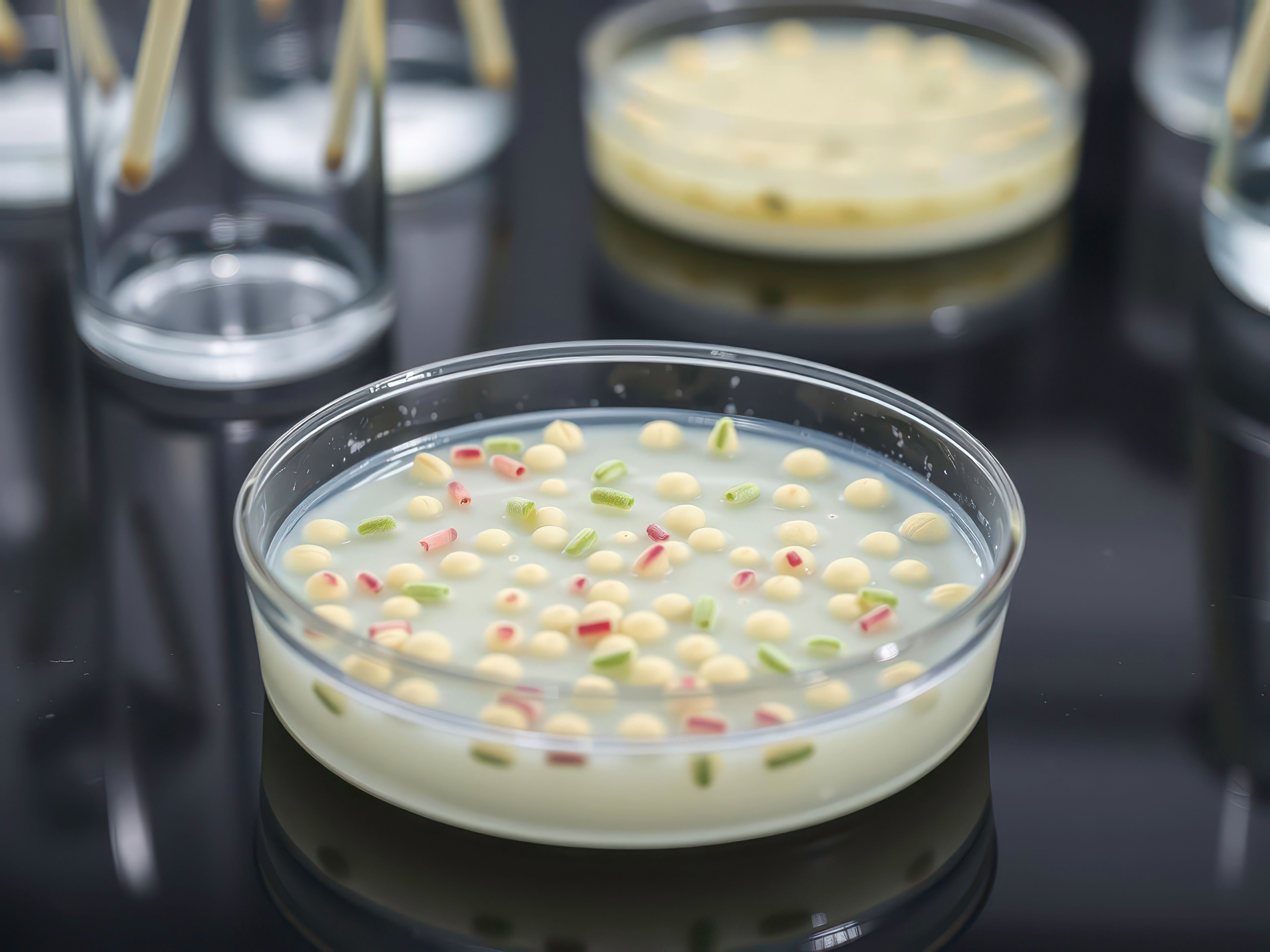
The current ongoing challenge, though, is that the traditional techniques often used for this purpose, which include mass spectrometry (MS) and chromatography, are often costly and inefficient (1). In this study, Zhao and the team attempted to improve on these traditional methods. Their approach leveraged the strengths of Raman spectroscopy and advanced deep learning models (2). Their work focuses on using optimized CNNs to process Raman spectra and accurately identify different types and batches of culture media (2).
To set up the experiment, the research team collected the Raman spectra from multiple samples of culture media using a confocal Raman spectrometer. Despite the samples exhibiting similar spectral features, subtle differences in peak intensities were detected (1). These spectral data were preprocessed and then input into three different machine learning models for training and validation: a principal component analysis with a support vector machine (PCA-SVM), an original CNN, and a structurally enhanced optimized CNN.
Then, the researchers compared the three models. Ultimately, they determined that the PCA-SVM model, which employed eight principal components capturing most of the data variance, was the second-best. It obtained a strong 99.19% accuracy and 98.39% precision (1). The original CNN model, on the other hand, performed poorly with an accuracy of only 71.89%, largely because of a limited training data set and insufficient model depth (1).
Therefore, because of the results achieved with the abovementioned methods, the optimized CNN model performed the best. This model achieved a perfect accuracy rate of 100% in distinguishing between various culture media types by incorporating batch normalization, max-pooling layers, and fine-tuning the convolutional parameters (1).
Once the best-performing model was determined, the team conducted external validation using unseen data from different media models and batches. The optimized CNN maintained its high performance, demonstrating strong generalization capabilities (1).
Overall, this study not only presents a model that can yield time and cost savings. The proposed method not only automates and accelerates the identification process, but it also reduces dependence on expensive instrumentation and expert interpretation (1).
Because the optimized CNN-based Raman spectroscopy model achieved a perfect identification rate and strong generalization performance, the researchers demonstrated how their method offers a promising new standard for ensuring consistency in culture media and other biological materials (1). As industries continue to seek scalable, automated, and reliable quality control methods, this study is the latest example of using machine learning (ML) with Raman spectroscopy to solve an ongoing challenge in pharmaceutical analysis.
References
- Wan, Y.; Jiang, Y.; Zheng, W.; et al. Rapid and High Accuracy Identification of Culture Medium by CNN of Raman Spectra. Spectrochimica Acta Part A: Mol. Biomol. Spectrosc. 2025, 329, 125608. DOI: 10.1016/j.saa.2024.125608
- Conduct Science, Culture Media: Classification, Types, and Relevance. Conduct Science. Available at: https://conductscience.com/culture-media/#:~:text=What%20Is%20Culture%20Media?,one%20type%20of%20culture%20media. (accessed 2025-04-09).


AI Boosts SERS for Next Generation Biomedical Breakthroughs
July 2nd 2025Researchers from Shanghai Jiao Tong University are harnessing artificial intelligence to elevate surface-enhanced Raman spectroscopy (SERS) for highly sensitive, multiplexed biomedical analysis, enabling faster diagnostics, imaging, and personalized treatments.


Artificial Intelligence Accelerates Molecular Vibration Analysis, Study Finds
July 1st 2025A new review led by researchers from MIT and Oak Ridge National Laboratory outlines how artificial intelligence (AI) is transforming the study of molecular vibrations and phonons, making spectroscopic analysis faster, more accurate, and more accessible.

Nanometer-Scale Studies Using Tip Enhanced Raman Spectroscopy
February 8th 2013Volker Deckert, the winner of the 2013 Charles Mann Award, is advancing the use of tip enhanced Raman spectroscopy (TERS) to push the lateral resolution of vibrational spectroscopy well below the Abbe limit, to achieve single-molecule sensitivity. Because the tip can be moved with sub-nanometer precision, structural information with unmatched spatial resolution can be achieved without the need of specific labels.

Machine Learning and Optical Spectroscopy Advance CNS Tumor Diagnostics
July 1st 2025A new review article highlights how researchers in Moscow are integrating machine learning with optical spectroscopy techniques to enhance real-time diagnosis and surgical precision in central nervous system tumor treatment.

AI and Dual-Sensor Spectroscopy Supercharge Antibiotic Fermentation
June 30th 2025Researchers from Chinese universities have developed an AI-powered platform that combines near-infrared (NIR) and Raman spectroscopy for real-time monitoring and control of antibiotic production, boosting efficiency by over 30%.

