Advancing Metabolite Identification with a Compact Infrared Ion Spectroscopy Platform
Key Takeaways
- IRIS combines MS sensitivity with IR-based structural fingerprints, overcoming MS limitations in metabolite identification.
- Recent advancements in compact, high-power systems have made IRIS more practical for industrial applications.
Metabolite identification is critical in drug development, with mass spectrometry (MS) as the primary tool, but limited in full structural elucidation. Infrared ion spectroscopy (IRIS) overcomes some of these limitations by combining MS sensitivity with IR-based structural fingerprints, enabling characterization without reference standards. Spectroscopy spoke to Giel Berden regarding applications in metabolite identification by determining the site of glucuronidation and phase I oxidation in selected drug molecules.
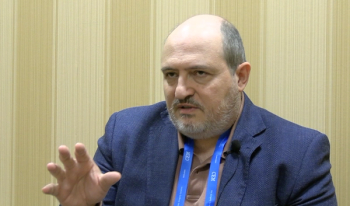
Metabolite identification is critical in drug development, with mass spectrometry (MS) as the primary tool, but limited in full structural elucidation. Infrared ion spectroscopy (IRIS) overcomes some of these limitations by combining MS sensitivity with IR-based structural fingerprints, enabling characterization without reference standards. Traditionally reliant on large-scale lasers, recent advances in compact, high-power systems have made IRIS more practical. Giel Berden (Radboud University and the High Field Magnet Laboratory and Free Electron Lasers for Infrared eXperiments [HFML-FELIX]) has developed for Johnson & Johnson a stable, small-footprint IRIS platform capable of reproducibly analyzing diverse metabolites, supporting its transition from academic research to routine industrial use. A recent study employed IRIS to differentiate isomeric compounds and demonstrated the robustness of a newly developed IRIS platform. Spectroscopy spoke to Berden, corresponding author of the resulting paper (1), showcasing applications in metabolite identification by determining the site of glucuronidation and phase I oxidation in selected drug molecules.
What are the main limitations of mass spectrometry (MS) in metabolite identification, and how can spectroscopy help overcome them?
MS is the analytical technique of choice for characterizing metabolites, particularly when combined with chromatographic methods. However, it frequently fails in complete structural elucidation, necessitating other orthogonal techniques, such as NMR or infrared spectroscopy. Spectroscopy provides information on resonances in molecules that are determined by the nature of chemical bonding within the molecules.
How does nuclear magnetic resonance (NMR) spectroscopy complement MS in drug metabolism studies, and what are the practical limitations of NMR?
NMR is an important tool in metabolism studies. Despite recent advances, several micrograms of purified analyte are still needed for successful structural elucidation. The complexity of biological matrices complicates the purification process, making it labor-intensive and time-consuming. In liquid chromatography ([LC])-MS, purification is less demanding or not needed at all, since the ion of interest can be mass-isolated from the complex mixture.
Explain the basic principle of infrared ion spectroscopy (IRIS) and how it differs from traditional infrared spectroscopy.
In traditional infrared absorption spectroscopy, sample density and optical pathlength can usually be adjusted such as to bring the attenuation of the infrared light into an observable range. However, this is not the case for the gaseous ions inside an MS; due to coulombic repulsion, densities are limited to values that are about 10 orders of magnitude below what is used for typical absorption experiments. Therefore, IRIS is not based on monitoring light attenuation. Instead, IRIS is a tandem MS (MS/MS) technique based on wavelength-dependent infrared photo-fragmentation of mass-selected ions inside an MS. The detection of fragment ions correlates with the absorption of infrared photons occurring when the frequency of the infrared laser is resonant with one of the vibrational transitions of the trapped ions. An infrared spectrum is then obtained by plotting the fragmentation yield as a function of the laser wavelength. It is interesting to note that the sensitivity of IRIS experiments is thus the same as for any other MS/MS experiment. Therefore, an infrared spectrum can be generated for any m/z peak having sufficient intensity for an MS/MS measurement.
In what ways does IRIS improve structural elucidation of metabolites, especially when MS/MS data alone are insufficient?
IRIS provides the infrared fingerprint of the metabolites. The presence or absence of vibrational bands in the infrared spectrum are connected to specific functional groups in the metabolite. For example, a band around 3600 cm-1 can easily be attributed to a free OH. Additionally, vibrational frequencies can be reliably predicted with quantum-chemical calculations, mitigating the need of reference compounds for structural elucidation.
Why is the 2800–3800 cm⁻¹ spectral range particularly useful in IRIS for organic molecule identification?
This spectral range covers the C-H, O-H, and N-H stretching vibrations that often provide enough detailed structural information so that small molecule identification is possible. Another very interesting range is 500-2200 cm-1 , which is currently only accessible at large-scale infrared free electron laser user facilities such as Radboud University’s FELIX facility. Turn-key, easy-to-use lasers for industrial applications of IRIS are not yet available at these long wavelengths.
Describe the role of density functional theory (DFT) in IRIS and how it aids in the identification of unknown metabolites.
One of the key advantages of using IRIS is that the IR spectra are obtained in the gas phase and therefore can be reliably calculated using DFT. This enables identification of compounds for which reference standards are not available. Even in the case that no definite identification can be obtained based on DFT alone, DFT will help to significantly narrow down the list of candidate structures, thereby substantially reducing the number of reference molecules that must be synthesized.
What is tagging spectroscopy in the context of IRIS, and what are its advantages and limitations?
In tagging spectroscopy, a weakly bound complex of the ion of interest with a tag (often N2) is dissociated instead of the ion itself. These complexes can only be formed in a cryogenic mass spectrometer. The advantage for IRIS is higher spectral resolution. Additionally, one does not need to dissociate the ion itself, so low-power infrared lasers can be used to remove the tag. A drawback is that cryogenic mass spectrometers are not yet available from commercial MS manufacturers.
Why is high laser power crucial for efficient IR multiple-photon dissociation (IRMPD), and what are the challenges when it's lacking?
The energy of an infrared photon is small compared to typical bond dissociation energies. Thus, it requires absorption of tens or even hundreds of photons to induce dissociation. High power lasers provide a large dynamic range for IRIS, which warrants the reliable detection of all vibrational bands, which is crucial for molecular identification. In other words, if the laser power is too low, you will miss several vibrational bands, which makes the identification process difficult or unreliable.
How can IRIS be used to distinguish between positional isomers of drug metabolites, and why is this important in pharmaceutical research?
The short answer is that positional isomers have different infrared fingerprint spectra and are therefore distinguishable with IRIS. In pharmaceutical research, drug metabolite identification is an essential part of the drug discovery and development process, where one identifies biotransformation products of drug molecules in the human body. Positional isomers can have significantly different effects in the body due to variations in their interactions with other molecules, leading to variations in their biological activity.
What technological developments have made IRIS more suitable for industrial applications, and what future improvements could expand its utility?
First, mass spectrometers with optical access to the trapped ion population are commercially available. These MS platforms are identical to the regular platforms found in industrial laboratories, with the same graphical user interface and data analysis software, ion sources, coupling to LC, and so on. Secondly, the development of infrared lasers. We tested several different types of infrared (IR) lasers, all commercially available, for their use in IRIS. We found that high-power, high-repetition-rate lasers outperform other lasers in photodissociation yield and that they offer advantages in terms of cost-effectiveness and practical implementation in an analytical laboratory not specialized in laser spectroscopy. The current IRIS platform located at Johnson & Johnson uses a commercial infrared laser that is fully integrated with the commercial MS. This IRIS setup is very easy to use and very stable: infrared spectra recorded over a timespan of one year are identical, without any re-adjustments or recalibrations made to the instrument. For the future, the development of high-power table-top lasers capable of reaching wavelengths down to 500 cm-1 (20 µm) allows observation of more vibrational bands, which would greatly facilitate the molecular identification process.
Reference
- van Wieringen, T.; Lubin, A.; van Outersterp, R. et al. Development of a Robust Platform for Infrared Ion Spectroscopy: A New Addition to the Analytical Toolkit for Enhanced Metabolite Structure Elucidation. Anal. Chem. 2025.DOI:
10.1021/acs.analchem.5c03593 - van Wieringen, T.; Lubin, A.; van Outersterp, R.; Martens, J.; van Beelen, E.; Oomens, J.; Cuyckens, F.; Berden, G. Development of a Robust Platform for Infrared Ion Spectroscopy: A New Addition to the Analytical Toolkit for Enhanced Metabolite Structure Elucidation. Anal. Chem. 2025.DOI:
10.1021/acs.analchem.5c03593
Newsletter
Get essential updates on the latest spectroscopy technologies, regulatory standards, and best practices—subscribe today to Spectroscopy.


