Carbon Quantum Dots Tested for Hypochlorite Detection and Cell Imaging
In a study led by scientists from Northwest University in Xi’an, China, carbon quantum dots were tested on how well they can detect hypochlorite and aid in cellular imaging.
In a study led by scientists from Northwest University in Xi’an, China, carbon quantum dots were tested on how well they can detect hypochlorite and aid in cellular imaging (1). The findings were published in Spectrochimica Acta Part A: Molecular and Biomolecular Spectroscopy.


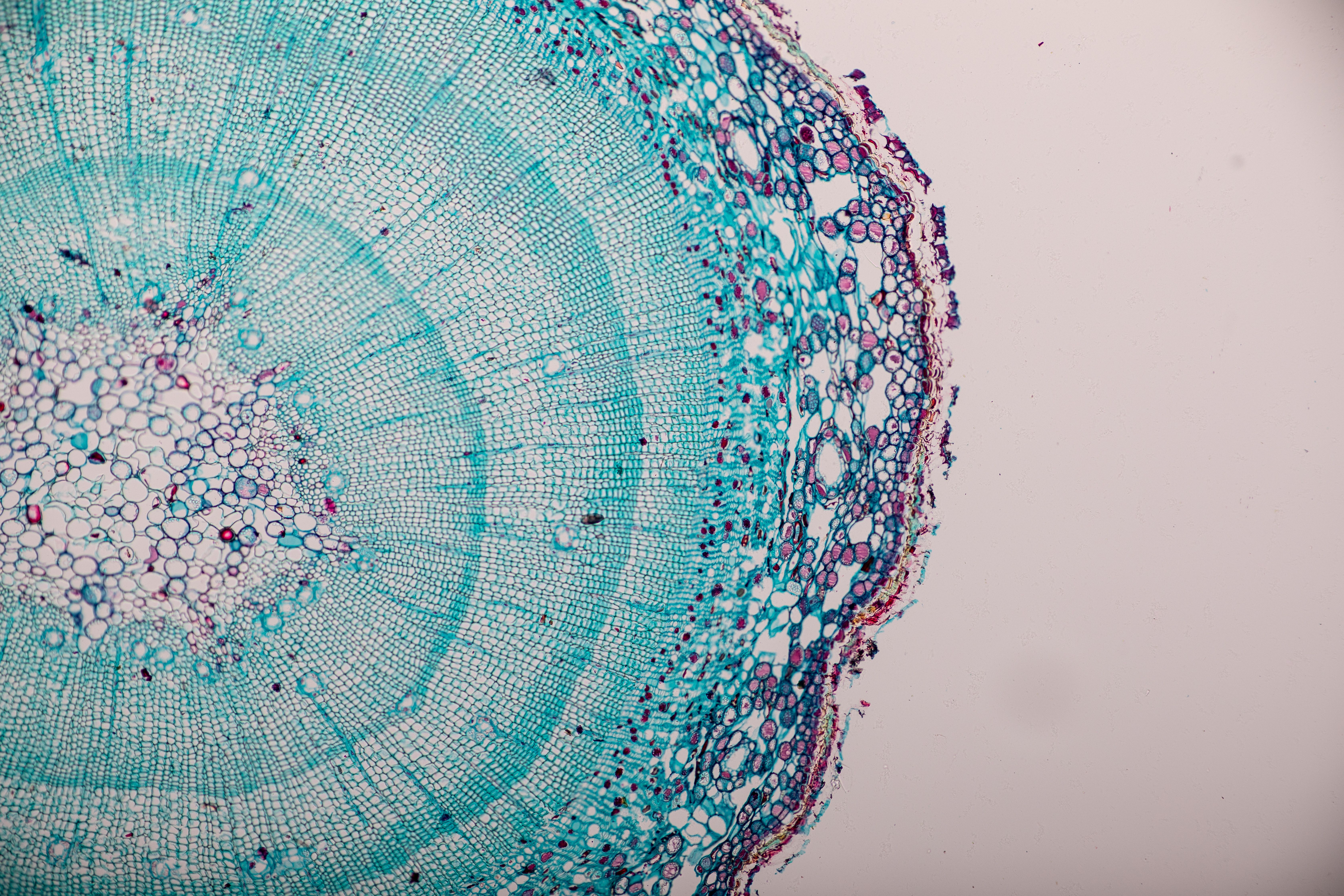
Cross-section Dicot, Monocot and Root of Plant Stem under the microscope for classroom education. | Image Credit: sinhyu - stock.adobe.com
Hypochlorite (ClO-) is seen as an important reactive oxygen species that helps in different physiological and pathological processes within organisms. It can be used as a disinfectant and preservative for environmental sanitization and food preservation. Unfortunately, excessive ClO- can disturb redox balances and cause severe threats to human health, like respiratory irritation, lung damage, atherosclerosis, and cancer. This has led to a need for accurate and sensitive ClO- concentration monitoring techniques, especially so regarding the fields of life sciences, food, and the environment.
There are various methods for analyzing ClO-, including high performance liquid chromatography (HPLC), surface-enhanced Raman scattering, colorimetry, and fluorescence methods. Of the above methods, fluorescence and colorimetry have shown high accessibility, easy preparation, non-invasive nature, high sensitivity, and good comparability for bio-samples. This led the scientists to choose dual-mode methods for potential and application prospects for analysis and detection.
As part of the experiment, bright green, fluorescent carbon quantum dots (G-CQDs) were synthesized using a one-step hydrothermal method, which has citric acid and acriflavine precursors. Carbon dots (CDs) are crystalline and “zero-dimensional” carbon nanomaterials that have gained widespread attention; green CDs are specifically synthesized from renewable biomass, such as plants and other organic biomaterials (2).
Read More: Green Carbon Dots: Synthesis, Characterization, Properties and Biomedical Applications
Multiple characterization procedures were used throughout this experiment. The morphology and size of G-CQDs were measured by transmission electron microscopy (TEM), while Fourier transform infrared spectroscopy (FTIR), X-ray photoelectron spectroscopy (XPS), and zeta potential characterization assisted in helping the G-CQDs illustrate a significant amount of uniformly dispersed –NH2 and –OH on the surface. The fluorescence and colorimetric analysis helped in displaying a wide linear range and a low detection limit response to ClO-.
After the main part of the experiment, G-CQDs were applied for visualizing and quantitively detecting ClO- in drinking water samples, with the results showing a satisfactory recovery rate. Additionally, the dots showed good water solubility, optical stability, and optimal biocompatibility, all of which foreshadow a promising analysis approach in cell imaging and ClO- detection in living cells. This experiment highlighted the benefits and potential of G-CQDs, showing their capabilities in ClO--associated disease prevention and early clinical diagnosis.
References
(1) Chen, H.; Li, D.; Zheng, Y.; Wang, K.; et al. Construction of Optical Dual-Mode Sensing Platform Based on Green Emissive Carbon Quantum Dots for Effective Detection of ClO and Cellular Imaging. Spectrochim. Acta Part A: Mol. Biomol. Spectrosc. 2024, 309, 123733. DOI: https://doi.org/10.1016/j.saa.2023.123733
(2) Jing, H. H.; Bardakci, F.; Akgöl, S.; Kusat, K.; et al. Green Carbon Dots: Synthesis, Characterization, Properties and Biomedical Applications. J. Funct. Biomater. 2023, 14 (1), 27. DOI: https://doi.org/10.3390/jfb14010027


LEGO Bricks: A New Standard for Evaluating Fluorescence in Raman Spectroscopy
July 24th 2024Researchers have proposed an innovative approach to tackling fluorescence interference in Raman spectroscopy by using LEGO blocks as standard samples. This new method offers a low-cost, rugged, and reproducible alternative to the complex liquid mixtures traditionally used in such studies, marking a significant advancement in the field of spectroscopic analysis.


