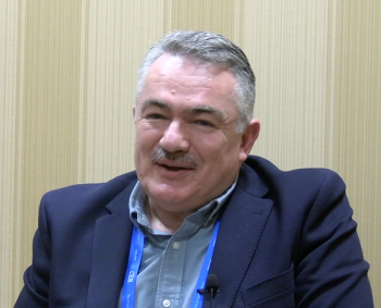
Improving Biomarker Detection with Surface-Enhanced Resonance Raman Scattering: An Interview with 2025 Charles Mann Award Winner Marc Porter (Part 1)
Spectroscopy speaks with Marc Porter, Distinguished Professor in Chemical Engineering at the University of Utah, for the first of a three-part conversation regarding his work involving the development and evaluation of a highly sensitive biomarker detection strategy using surface-enhanced resonance Raman scattering (SERRS), which offers stronger signal amplification than traditional surface-enhanced Raman scattering (SERS). Porter will receive the 2025 Charles Mann Award, presented to an individual who has demonstrated advancement(s) in the field of applied Raman spectroscopy.
The laboratory of Marc Porter, Distinguished Professor in Chemical Engineering at the University of Utah, is probably best known for investigating applications of surface-enhanced Raman scattering (SERS) as a read-out tool in multiplexed human and animal health care testing. Recent work involved the development and evaluation of a highly sensitive biomarker detection strategy using surface-enhanced resonance Raman scattering (SERRS), which offers stronger signal amplification than traditional SERS. Focusing their research on the detection of mannose-capped lipoarabinomannan (ManLAM), a biomarker for active tuberculosis (TB) infection, the laboratory’s findings suggest that SERRS-based immunoassays could significantly improve low-level biomarker detection, enhancing disease diagnostics and patient outcomes.
Porter will receive the 2025 Charles Mann Award, presented to an individual who has demonstrated advancement(s) in the field of applied Raman spectroscopy. The award will be presented at SciX 2025, taking place from October 5 to 10 at the Northern Kentucky Convention Center in Covington, Kentucky. Spectroscopy spoke to Porter about this study and the resulting article (1) as part of our continuing interview series with SciX award winners.
What motivated the shift from using surface-enhanced Raman spectroscopy (SERS) to surface-enhanced resonance Raman spectroscopy (SERRS) in your biomarker detection platform?
We all know that early disease detection goes conjointly with timely intervention and, as a result, improved treatment outcomes. In vitro diagnostic tests play an important role throughout this process. These tests aid in the diagnosis of a disease, guide treatment decisions, and monitor response to treatment. Some tests also can identify if a patient has an increased level of risk in developing diseases like breast cancer and Alzheimer’s disease.
Health care providers gauge the utility of a diagnostic test on metrics like clinical sensitivity and clinical specificity. Both measures of performance are directly linked to analytical figures of merit like analytical sensitivity, limit of detection, limit of quantitation, accuracy, and precision. The motivation to examine the utility of SERRS as an approach to biomarker readout in our immunoassay based on gold nanoparticle labeling was driven by the significant levels of improvements in limits of detection and analytical sensitivities reported by several research groups, including those from past and likely to be future Mann Awardees. In a few instances, SERRS produced signal intensities that rivaled those of fluorescence, but with spectral features that have a much higher level of molecular content and much narrower widths that enable a greater level of multiplexing.
Can you explain how resonance Raman effects enhance signal strength compared to conventional SERS?
Surface-enhanced resonance Raman scattering (SERRS) couples the ~106 enhancement in signal strength of SERS with that from resonance Raman scattering (RRS). RRS scattering occurs when the wavelength for excitation overlaps with energy gap of a dipole-allowed electronic transition of a molecule. Raman active vibrational modes that are efficiently coupled to the electronic transition will exhibit intensity enhancements by factors of 102-106 times greater than those for normal (unenhanced) Raman scattering. Recent work has shown the SERRS responses can rival and, at times, surpass those for measurements based on fluorescence. Let me add that our immunoassay platform, which mimics the nanoparticle-on-mirror architecture first proposed by Metiu and examined later by Moskovits, incorporates the localized plasmon resonance of gold nanoparticles with the surface plasmon polariton of an underlying gold mirror. A more detailed discussion of the structure of our immunoassay architecture is discussed later in the interview.
How did you address the challenge of optimizing the density and distributions of the nanoparticle labels in your SERRS immunoassay?
The questions about optimizing nanoparticle label density and distributions are related to efficiencies in ability to capture the antigen and then label it after capture. For perspective, let me begin my response by describing the layout of our sandwich-styled immunoassays based on SERS and then detail how we have modified its architecture for SERRS. The layout embodies several of the design elements common to ELISA, including a substrate coated with a layer of antibodies for the selective capture of the target antigen and a selective labeling step that is coupled to a signal amplification mechanism. Our layout, however, features two distinct differences. First, the capture address is a gold film modified with a layer of capture antibodies. Second, the label for captured antigen consists of a gold nanoparticle core coated with a monolayer of an intrinsically strong Raman reporter molecule (RRM) and then a layer of tracer antibodies. We refer to the modified gold nanoparticles are as extrinsic Raman labels (ERLS). This design takes advantage of the large enhancement in signal strength gained from SERS as a basis to compete, if not surpass, the detection capabilities of enzyme-linked immunosorbent assay (ELISA), while avoiding the time and temperature required for enzymatic signal generation. The layout for our SERRS immunoassays closely parallels that of the design for SERS, but has, one notable difference. For SERRS, the RRM is coated on the capture substrate and not on the antibody-coated nanoparticles employed to tag captured antigens (the “why’ for this change will be discussed shortly). These designs have applied to the detection of a wide range of biomarkers for disease, toxins, viruses, and bacteria, with the goal of “taking health care testing to the patient.”
The concerns over label density and distribution arose when measuring the nanoparticle distributions and densities from atomic force microscopy (AFM) images for samples prepared for SER readout and for SERRS readout, which, as described above, were prepared differently. A comparison of particle densities for samples exposed to the same ManLAM concentrations yielded densities that were ~35% higher for the SERRS samples with respect to the SERS samples at all the ManLAM concentrations tested (1-50 ng/mL). This difference accounts for a small portion of the larger slope (10´) of the calibration curve for SERRS versus that for SERS. The intriguing aspect of this observation is that the measure densities are more about four orders of magnitude below that for a closest packed layer of particles with a 60 nm diameter, which point to the possibility of markedly improving the detection power of both SERS and SERRS. Experiments to test potential pathways to improve the densities and distributions, for example, by changing the proportions of the thiolated Cy% and DSP in the self-assembly mixture will begin soon. More to follow soon!
What makes mannose-capped lipoarabinomannan (ManLAM) a particularly useful biomarker for active tuberculosis detection?
Tuberculosis has killed more people than any other infectious disease in human history. The World Health Organization (WHO) estimates that there were 10.8M people newly infected with TB in 2023 and 1.25M associated deaths. Most of this ongoing mortality occurs in low- and middle-income countries and is strongly linked to the fact that millions of newly infected TB victims go undiagnosed each year from the lack of an accurate and affordable PON TB test.
The value of developing a field-deployable, accurate, low-cost TB test in underscored by the fact that TB can be cured if detected early in its progression but suffers a case fatality rate of ~50% if left untreated. While there are several tests that have been ingeniously adapted to meet this need, the associated costs remain out of reach for many of those who need it.
ManLAM has the potential to fill a long-standing, global need for biomarkers that can be reliably used to detect tuberculosis (TB). ManLAM has been found in the urine and serum of individuals infected with Mycobacterium tuberculosis (Mtb), the causative agent for tuberculosis (TB). ManLAM is a virulence factor associated with the infectious pathology of tuberculosis (TB). It is a unique and highly branched lipoglycan (~17.3 kDa) component of the organism’s cell wall (Mtb) and is up to 15% of the mass of the organism. This collection of properties and characteristics has made it an intriguing candidate as a TB biomarker in clinical settings and in point-of-care settings in field-portable test kits. There are several research groups around the world, including those we have collaborated with at Colorado State University and Rutgers University, working to make this happen.
In your experiments, how did you validate the 10× improvement in the limit of detection and the 40× increase in sensitivity?
We typically carry out a brief assessment of the performance of a diagnostic test in its early stages of development with the two most used analytical figures of merit: limit of detection (LoD) and analytical sensitivity, the latter of which is also a measure of the linearity of a calibration curve. These metrics are determined using the data from three to seven replicate assays.
The next steps in this assessment were put on hold while we worked on needed improvements in our approach to pretreating serum and urine samples prior to analysis. These improvements, which include combating the impact of the sample matrix with a user-friendly approach to sample pretreatment, will be discussed later in the interview. Once we lock down the sample preparation methodology, we will focus on furthering our assessment of the analysis of ManLAM by SERRS by testing the specificity and robustness of measurement against interferences that degrade the effectiveness of our sample pretreatment method and how small changes in operating parameters like temperature and pH may have an impact on the measurement. We also task resources to gain an understanding of the nature of the sample matrix. We may, for example, determine the range of protein concentrations and viscosity in each patient cohort. Both parameters, which can alter binding reaction rates, can undergo increases or decreases depending on the nature of the disease and the physiological response of the patient. This phase of the effort will also carry out a much more exacting assessment on the limit of detection (LoD), and the accuracy and precision of the measurement. All these tasks will be interwoven with an extensive series of double-blinded cohort studies to determine the clinical accuracy of our test and to identify any necessary refinements.
Join us tomorrow for the second part of this interview. Discover how thiolated-Cy5 dyes and gold capture surfaces are revolutionizing detection sensitivity in SERRS-based immunoassays. Leveraging the powerful quenching and resonance-enhancing properties of Cy5 on gold surfaces, this innovative platform achieves single-molecule sensitivity, outpacing traditional methods like ELISA and PCR. Learn how this technique enables faster, more reliable detection in point-of-need settings, and explore its potential for real-world clinical deployment. From rugged portable Raman spectrometers to pioneering vertical flow assay integration, the future of diagnostics is here, powered by advanced plasmonics and fluorescence engineering.
References
1. Owens, N. A.; Pinter, A.; Porter, M. D. Surface-Enhanced Resonance Raman Scattering for the Sensitive Detection of a Tuberculosis Biomarker in Human Serum. J. Raman Spec. 2019, 50 (1), 15-25. DOI:
Newsletter
Get essential updates on the latest spectroscopy technologies, regulatory standards, and best practices—subscribe today to Spectroscopy.


