SPEC PerkinElmer 11.10
News|Articles|March 28, 2025

New Collaboration Between Thermo Fisher and the Chan Zuckerberg Institute Aims to Improve Cryo-Electron Microscopy Technology

Author(s)Will Wetzel
Fact checked by: Jerome Workman, Jr.

Thermo Fisher Scientific Inc., a global leader in scientific solutions, announced in a press release that they have secured a Technology Alliance Agreement with the Chan Zuckerberg Institute for Advanced Biological Imaging (CZ Imaging Institute) (1). This partnership is designed to help spur advancements in cryo-electron microscopy (cryo-EM) technology, enhancing researchers' ability to visualize human cells at unprecedented resolution (1). The long-term goal of this partnership is to lead to new therapeutic developments and scientific discoveries (1).

At its core, cryo-Em is an advanced imaging technique (2). Scientists use this technique in order to better understand biological molecules (2). Because of the technique’s core focus, scientists often use cryo-EM technology in disease research and drug development, and it also plays a role in developing new vaccines (1,2). Currently, although cryo-EM technology has improved, limitations still remain. For example, researchers recognize the need for improved imaging contrast and resolution (1).

This partnership between Thermo Fisher Scientific and the CZ Imaging Institute will focus on advancing laser phase plate technology to refine the capabilities of cryo-EM technology, which will allow scientists to explore the inner workings of human cells in three dimensions at nanoscale resolution (1).

A key aspect of this effort builds on previous work with Professor Holger Müller and his team at the University of California, Berkeley, and Lawrence Berkeley National Laboratory (Berkeley Lab). The collaboration, which has already led to the invention and ongoing development of the continuous laser phase plate for electron microscopy, will now be expanded with Thermo Fisher and the CZ Imaging Institute working together to further refine the technology (1). By enhancing imaging precision, this partnership will enable breakthroughs in biological research and medical science (1).

“Broadening our understanding of basic cellular biology has the potential to unlock new layers of scientific research and therapeutic development,” Trisha Rice, vice president and general manager of life sciences for Thermo Fisher’s electron microscopy business, said in a press release (1). “We are excited to collaborate with the CZ Imaging Institute to create new approaches in analytical science that aid future discovery.”

This initiative aligns with the broader mission of the Chan Zuckerberg Initiative (CZI) to tackle major scientific challenges. A central goal of CZI is to develop advanced imaging systems that provide extraordinary insights into human biology (1). By leveraging the expertise of scientists and cryo-EM specialists from both organizations, the partnership will maximize the impact of laser phase plate technology, allowing researchers worldwide to gather more comprehensive data and advance cutting-edge biological research (1).

Matthias Haury, acting executive director and chief operating officer of the CZ Imaging Institute, highlighted the collaboration’s significance.

“At the Imaging Institute, we’re focused on innovating breakthrough imaging technologies to enable scientists to see and measure what is currently invisible within our cells and gain deeper insights into the biological mechanisms of human health and disease,” Haury said in a press release (1). “We want to advance not only the experimental side of imaging science but also deliver solutions to the community. Technology alliances with industry partners like Thermo Fisher allow us to accelerate and scale technology development and share these advances with scientists worldwide more rapidly.”

The CZ Imaging Institute actively works on developing imaging hardware and software tools to create multimodal biological imaging systems that enhance scientific understanding (1). Thermo Fisher Scientific Inc.’s mission is to support customers in making the world healthier, cleaner, and safer by accelerating life sciences research, addressing complex analytical challenges, increasing laboratory productivity, or advancing patient care through diagnostics and therapeutics (1).

With this partnership, Thermo Fisher Scientific and the CZ Imaging Institute will have an opportunity to align both organization’s goals with the broader mission of advancing scientific discovery.

References

  1. Thermo Fisher Scientific, Thermo Fisher Scientific and Chan Zuckerberg Institute for Advanced Biological Imaging Collaborate to Further Understanding of Human Cells. Thermo Fisher Scientific. Available at: https://newsroom.thermofisher.com/newsroom/press-releases/press-release-details/2025/Thermo-Fisher-Scientific-and-Chan-Zuckerberg-Institute-for-Advanced-Biological-Imaging-Collaborate-to-Further-Understanding-of-Human-Cells/default.aspx (accessed 2025-03-27).
  2. Carreras, H. Z. Technology Network. Available at: https://www.technologynetworks.com/analysis/articles/cryo-electron-microscopy-principle-strengths-limitations-and-applications-377080#:~:text=Cryo%20EM%20is%20an%20advanced,the%20sample%20from%20these%20projections. (accessed 2025-03-27).


Related to this article

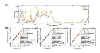
Monitoring Sugar Consumption and Ethanol Production During Yeast Fermentation Using a Raman Plate Reader and Multivariate Analysis
This column shows that a Raman plate reader paired with partial least squares regression can simultaneously quantify glucose, fructose, sucrose, and ethanol in yeast fermentation samples, using turbidity-matched calibration standards to correct for yeast-induced turbidity, and offers a rapid, label-free, high-throughput method for tracking sugar consumption and ethanol production in strain screening, media optimization, and process development.
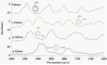
Highly Substituted Benzene Rings—The Rest of the Story II: Spectra
This column, the second installment on highly substituted benzene rings, shows that tri- and tetra-substituted substitution patterns can be distinguished by whether the ring bending peak is present and by the position of the in-phase aryl C-H wagging peak, which shifts according to how many adjacent hydrogens the ring carries.
Healthcare administrator reviewing an insurance policy schedule with a stethoscope on a clipboard, health insurance coverage, medical underwriting, prior authorization, and patient claims processing. | Image Credit: Joi_studio - stock.adobe.com
In a SciX interview, Marc Porter covered a lot of ground, from a student-driven pivot into immunoassays to a baseball analogy for Stokes and anti-Stokes scattering, and shares the open questions he is still chasing.
Ingots of pure copper or pink gold on a black background. | Image Credit: Phawat - stock.adobe.com
At SciX, Kevin Wilkinson of the Universite de Montreal explains how faster data acquisition and new software have cut analysis time and how a particle’s chemical composition can link arsenic-bearing emissions back to a copper refinery’s processes.
Spectral Frontiers: Seeing the Unseeable Microspectroscopy
Microspectroscopy, the marriage of spectroscopic chemical identification with microscopic spatial resolution, has advanced from Perkin-Elmer's first commercial infrared microspectrometer in 1953 to today's nanoscale AFM-IR probes, synchrotron-brightness infrared beamlines, and compact hyperspectral tissue imaging devices being developed for AI-assisted cancer detection.