Key Points
- In the SERRS immunoassay, Cy5’s fluorescence is quenched upon adsorption to gold, removing background noise while enabling resonance enhancement. The thiolated form of Cy5 also facilitates strong and specific immobilization to gold surfaces through Au–S bonding, allowing precise antibody attachment and enhanced signal amplification.
- SERRS immunoassays demonstrate dramatically lower limits of detection (e.g., 2.3 pg/mL for MMP-7 vs. 31.8 pg/mL via ELISA) and benefit from sharper spectral features, reduced photobleaching, minimal background fluorescence, and single-laser multiplexing. These advantages position SERRS as a next-generation diagnostic platform.
- Development efforts are focused on stabilizing reagents (e.g., lyophilization), ruggedizing portable Raman units, and integrating with vertical flow assay (VFA) formats. Real-world deployment also requires assay standardization, regulatory compliance, training, and safe field practices—all critical for TB, cancer, and potential “Disease X” applications.
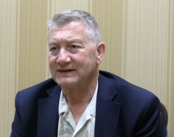
This is the second part of our interview with Marc Porter, Distinguished Professor in Chemical Engineering at the University of Utah and the recipient of the 2025 Charles Mann Award, presented to an individual who has demonstrated advancement(s) in the field of applied Raman spectroscopy.
Here, discussing his recent paper on the subject (1), Porter speaks on how thiolated-Cy5 dyes and gold capture surfaces are revolutionizing detection sensitivity in surface-enhanced resonance Raman spectroscopy (SERRS)-based immunoassays. Leveraging the powerful quenching and resonance-enhancing properties of Cy5 on gold surfaces, this innovative platform achieves single-molecule sensitivity, outpacing traditional methods like ELISA and PCR.
The Charles Mann Award will be presented at SciX 2025, taking place from October 5 to 10 at the Northern Kentucky Convention Center in Covington, Kentucky. Spectroscopy spoke to Porter about this study and the abovementioned article as part of our continuing interview series with SciX award winners.
Could you elaborate on the role of thiolated-Cy5 and the significance of the gold capture surface in your assay design?
Cy5 is a water-soluble, far-red fluorescent dye used as a label in high-sensitivity microscopy, flow cytometry, and nucleic acid assays. With an absorption maximum at 649 nm, it is well-suited for excitation by HeNe lasers at 633 nm and krypton and diode lasers at 647 nm. Note the use of Cy5 in SERRS takes advantage of two attributes. First, when adsorbed on gold, silver and many other metals, the dye’s fluorescence is quenched by non-radiative energy transfer mechanisms. This process eliminates a detectable fluorescence background that competes with the spectral features of Cy5 that are resonance enhanced. Second, resonance enhancement is enabled, i.e., turned on, when a gold nanoparticle that is coated with a tracer antibody tags a captured antigen. Upon irradiation at 633 nm, the nanometrically small gap that had formed between the gold nanoparticle and the underlying gold surface couples the enhancement from SERS and that of RRS. This coupling translates to notable improvements in LOD and analytical sensitivity in the SERRS-based immunoassay for ManLAM discussed earlier in this interview.
The use of a thiolated Cy5, which was prepared by the reaction of the N-hydroxysuccinimidyl ester of Cy5 with 2-aminoethanethiol, serves a second purpose. The sulfhydryl group at the end of a flexible chain, which is attached to Cy5’s extended p-electron system, can be used to immobilize the dye to gold surfaces by the formation of gold-bound thiolate, which cleaves the S-H bond. After purifying the product from the dye’s modification, capture substrates were prepared by first forming a mixed thiolate using an ethanolic mixture of thiolated Cy5 and dithiolbis (succinimidyl propionate), which was used to tether capture antibodies to the surface of the gold substrate by the reaction of DPS’s NHS ester to the sterically accessible amine groups located at the outer surface of the antibodies. The formation of a gold-bound thiolate is supported by the presence of a weak gold-sulfur stretch, ns(Au-S), at ~320 cm-1 in the SERRS spectrum of this system.
What are the main analytical advantages of SERRS-based immunoassays over existing TB diagnostic techniques like ELISA or polymerase chain reaction (PCR)?
The driver behind developing and testing SERS and SERRS as immunoassay readout tools was based the first reports demonstrating single molecule detection using both techniques. These findings led us to hypothesize that designing an immunoassay using SERS and SERRS as readout tools had the potential to surpass the detection capabilities of ELISA for biomarkers without the time and temperature requirements for enzyme activation and substrate turnover. The SERS and SERRS architectures for these tests described earlier in the interview are the results of this effort and involved replacing the enzymatic amplification processes operative in ELISA with plasmonic enhancements of SERS or the plasmonic and resonance Raman enhancements of SERRS.
During the early stages of development, we identified other advantages in using SERS and SERRS as a readout tool with respect to those for ELISA. These include:
- The optimum excitation wavelength for SERS is dependent on nanoparticle size, shape, and composition. Only one laser excitation wavelength is required for multiple analytes.
- The widths of the Raman spectral features for both SERS and SERRS are typically 10-100 times narrower than those of fluorescence. This characteristic minimizes the potential for overlap in the response from different molecular labels and is a key ingredient to the work on multiplexed analysis being pursued by research teams around the world.
- SERS using large diameter AuNPs (for example, 60 nm) requires excitation with red or longer spectral wavelengths. This strongly minimizes, if not virtually eliminates, the fluorescence interference of most biological media when at short wavelength excitation.
- Raman scattering is not sensitive to humidity or affected by oxygen or other quenchers. This characteristic facilitates applications in a variety of environments.
- SERS signals are less prone to photobleaching. This results in a more reliable and reproducible signal, adding the option to signal average longer if needed to further lower the LOD.
We recently validated our hypothesis in work focused on the detection of two well-established biomarkers for pancreatic cancer: serum carbohydrate antigen 19-9 (CA 19-9) and matrix metalloproteinase-7 (MMP-7). CA19-9 is the only FDA-approved biomarker for pancreatic cancer diagnosis, but it has a low level of specificity due to its upregulation by conditions not directly linked to the onset of pancreatic cancer. The LODs determined using our platform to analyze spiked human serum were 2.3 pg/mL for MMP-7 and 34.5 pg/ mL for CA 19-9. Tests using the same antibody pairs in ELISA yielded LODs of 31.8 pg/mL and 987 pg/mL for MMP-7 and CA 19-9, respectively. We also ran a set of analyses using three deidentified serum specimens from patients with pancreatic cancer. These results showed differences between the two types of measurements for MMP-7 of ~30% or less, whereas those for CA 19-9 differed in one instance by ~40%, but were not detectable on the other two samples. Taken together, these results point to the utility of these techniques for the analysis of pancreatic cancer biomarkers and, more broadly, to real-world samples.
What considerations went into adapting the SERRS platform for point-of-need (PON) diagnostic settings?
Adaption of the platform for PON applications has proceeded in parallel with that of our work for its use in clinical settings. It is important to note that almost all the tasks required for movement to the clinic will need to be incorporated into the adaptations of the platform for PON applications. That said, there are development and standardization considerations tied specifically to movement to the PON arena, which include the care and feeding of the portable Raman spectrometer, reagent stability, and safe practices. A few details on how we are addressing these needs follow below:
- Spectrometer: Establish the spectrometer's ruggedness (drop resistance and temperature susceptibility), portability (weight and size), operational time between recharge cycles concerning usage and recharge requirements, and optical accessories and adaptors for sample readout.
- Reagent stability: Develop and quantify the effectiveness of approaches to break the cold chain by lyophilizing antibodies and other time and temperature-sensitive reagents, and approaches for their preparation for use at the work site.
- Safe practices: Develop procedures and train manuals and videos to ensure the safety of field technicians and other end users and require all users to successfully complete all mandatory training exercises (laser safety, sample collection and preparation), and waste disposal) and demonstrate then demonstrate operation proficiency. Related items can be found in Hopkins’ recent publication (2).
What steps are needed to move this detection strategy from a research lab environment to widespread clinical use?
Moving our Raman-based immunoassay from the R&D laboratory to clinical settings entails several interconnected tasks. Each of these tasks focus on implementing the changes required to transform a proof-of-concept analysis system into a robust, reliable, and clinically validated diagnostic tool. A same sampling of these tasks include:
- optimizing and standardizing all assay procedures, including those related to collection, storage, and preparation of samples;
- defining instrument operational setting and standardize tests to determine, for example, if the laser’s output intensity is in or out of compliance with during deployment; and
- validating the clinical accuracy of the test, the data analysis software, data interpretation, and measurement levels for yes/no cutoffs for screening-based tests.
- developing safe and secure modes for short- and long-term data storage and transmission.
We have also worked to establish strong connections with our clinical colleagues and partners to gain clear perspective and insights for the specifications required for clinical acceptance, which include ease of use, regent preparation, and the footprint of the hardware and the utility connections required for operation.
Are there any foreseeable challenges in integrating this SERRS assay into vertical flow-style diagnostic devices?
As you know, other research groups are working to combine the detection strengths of SERS with the ability to markedly reduce the time required to complete the antigen capture and labeling steps in a vertical flow assay (VFA). VFAs slowly pass the liquid sample through a membrane-like material that is modified to act as a capture substrate. Several of these studies use porous materials impregnated with antibody-modified gold nanoparticles for this purpose. We have also been investigating the merits of integrating the two technologies in pursing the development a POC screening test for alphafetoprotein in human serum. This 70 kDa protein is an at-risk marker for hepatocellular carcinoma (HCC) and is part of a broader effort to develop a multiplexed panel screen to detect the early onset of this devastating disease. Our work with VFAs takes advantage of our extensive experience in developing a water quality for the potable water systems in the International Space Station (ISS) that used colorimetric-solid phase extraction (C-SPE). This technology was used to quantify biocide levels by impregnating porous, flow-through media with selective colorimetric indicators, which were interrogated by using a hand-held and microgravity certified diffuse reflection spectrometer and has been operational on ISS for more than a decade.
We view adapting our SERRS-based immunoassay to operate in a vertical flow format is one of the steps that will prove pivotal in the successful movement of ManLAM assay to function as a POC test for TB. This perspective, supported by our own experiences and by a literal explosion of work on VFAs around the world. Recent work has for example, demonstrated the ability to complete a vertical flow assay in spiked human serum within a few minutes. Other reports have been able to reach LODs for a mycotoxin at ~8 fg/mL. These developments, when coupled with strategies to carry out multiplexed biomarker detection, tell us to get these plans moving forward yesterday.
Join us tomorrow for the final part of this interview. Picture a world where diagnosing deadly diseases is as easy as pulling out a handheld device. With portable Raman spectrometers entering decentralized healthcare, even the most remote settings can benefit from instant analysis—slashing turnaround times and costs while boosting patient care. This innovation extends to our SERRS-based immunoassays: highly flexible, multiplexable, and ready for a range of biomarkers and pathogens. From TB to pancreatic cancer, these tools hold the promise of real-world impact, especially in TB-endemic regions where accurate, affordable, point-of-need tests could save millions. But that’s not all…emerging nanotechnologies and AI-driven breakthroughs are poised to redefine speed, sensitivity, and selectivity, bringing personalized, real-time diagnostics within reach. The future of global health is decentralized, powerful, and closer than ever, powered by Raman, SERRS, and unstoppable innovation.
References
1. Owens, N. A.; Pinter, A.; Porter, M. D. Surface-Enhanced Resonance Raman Scattering for the Sensitive Detection of a Tuberculosis Biomarker in Human Serum. J. Raman Spec. 2019, 50 (1), 15-25. DOI: 10.1002/jrs.5500
2. Hopkins, A. Overcoming Struggles in Implementing Handheld Raman Spectroscopy Across the Manufacturing Line. Spectroscopy 2017, 32 (6) 40-43.


