Submicron IR Detects and Localizes Microplastics in Biological Samples
Key Takeaways
- O-PTIR overcomes key constraints of FT-IR, Raman, and Py-GC-MS by delivering label-free, submicron chemical imaging in situ without contact, digestion, or tissue architecture disruption.
- PTFE in tonsil was identified via characteristic 1219/1159 cm⁻¹ bands and database matching (HQI >90), then localized by single-frequency and ratioed imaging to discrete ~1 µm regions.
This paper demonstrates the use of optical photothermal infrared (O-PTIR) spectroscopy for the detection, identification, and localization of microplastics in tissue samples.
Detecting and identifying microplastics in biological samples remains a significant analytical challenge, particularly for particles in the small micron and submicron range. Conventional techniques provide chemical identification but are limited in spatial resolution, sensitivity, or require destructive sample preparation. There is a clear need for analytical approaches that enable nondestructive, high-resolution chemical analysis directly within complex biological matrices. This paper demonstrates the use of optical photothermal infrared (O-PTIR) spectroscopy for the detection, identification, and localization of microplastics in tissue samples. Using representative biological examples, O-PTIR enables submicron spatial resolution imaging and chemical characterization without labeling or sample damage. These results highlight the significance of O-PTIR as an analytical measurement technique for studying microplastic bio-uptake and distribution within biological systems.
Microplastics (MPs) are polymeric particles with sizes between 1 µm–5 mm, while nanoplastics are generally defined as particles less than 1 µm. For simplicity, the term “microplastics” is used throughout this work to refer collectively to both microplastics and nanoplastics. MPs have been found to be ubiquitous in the environment, and recent publications report their presence in human organs such as the liver,¹ placenta,² and lung.³ It has also been reported that MPs can act as carriers of contaminants.4-5
While public awareness of MPs has increased, their effects on biological systems, including human cells and tissues, remain to be fully investigated. Among the wide range of particle sizes, smaller MPs (< 20 µm) are more likely to penetrate tissue and potentially cause harm. However, detecting and localizing these smaller particles in biological samples without prior digestion or sample damage remains challenging.
A variety of analytical techniques have been used for MP detection and identification, including Fourier transform infrared spectroscopy (FT-IR), Raman spectroscopy, and pyrolysis gas chromatography–mass spectrometry (Py-GC-MS).6,7 These methods provide high confidence in chemical identification but present limitations for in situ biological analysis. Conventional FT-IR is limited in spatial resolution, ATR-FTIR requires contact with the sample, Raman spectroscopy can suffer from low sensitivity and fluorescence interference, and Py-GC-MS is destructive and subject to interference from biological matrices.
O-PTIR has seen growing adoption by scientists across different fields over the years8 since its introduction and publication in 2016.9 Within the microplastics research community, the technique has been used to detect microplastic particles in a range of nonbiological environments, including snow,10 the atmosphere,11 and intravenous fluid delivery systems.12 In addition, researchers have shown that combining both IR and Raman modalities can improve the accuracy of MP identification.13
O-PTIR spectroscopy provides submicron spatial resolution in a noncontact and nondestructive mode. O-PTIR employs a pump–probe scheme in which a visible probe laser detects the photothermal response generated by a pulsed mid-infrared laser (Figure 1). The probe laser can also function as a Raman excitation source, enabling simultaneous acquisition of IR and Raman spectra from the same location. Fluorescence imaging can be integrated to guide measurements (Figure 2). In this work, O-PTIR and colocated fluorescence imaging are applied to detect, identify, and localize microplastics in tissue samples. A more detailed description of O-PTIR can be found
Materials and Methods
Tissue samples were prepared for analysis using standard histological approaches. Tonsillar tissue obtained from a patient was cryo-sectioned into 10 µm slices, washed, mounted onto CaF₂ windows, and allowed to dry prior to analysis. Periprosthetic tissue from a failed arthroplasty case was similarly prepared for spectroscopic measurements.
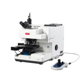
O-PTIR measurements were performed using the mIRage-LS sub-micron IR microscope (Photothermal Spectroscopy Corp, Santa Barbara, CA). Spectral acquisition was performed by sweeping the mid-infrared laser across the spectral range. Single-wavenumber imaging was conducted by tuning the laser to characteristic absorption bands and scanning the region of interest. Ratioed imaging was used to enhance contrast between polymer-specific and tissue-specific spectral features. Step sizes for imaging ranged from 50–250 nm, depending on the measurement.
The 532 nm visible probe laser can also be used as a Raman excitation source, enabling simultaneous acquisition of infrared and Raman spectra from the same location.13 Colocated fluorescence imaging was used in some cases to guide the identification of regions of interest.14,15
Database searching was undertaken with the Wiley KnowItAll database.
Results
Detection of Microplastics in Tonsillar Tissue
Tonsillar tissue sections were analyzed using O-PTIR. The brightfield image (Figure 3, top) did not reveal any distinct features that suggest the presence of microplastics. Random sampling of the tissue using O-PTIR suggests that the spectra can be categorized into two distinct groups (red and blue spectra in Figure 3, bottom). One group aligns with typical tissue spectra (1658 cm-1 and 1546 cm-1 for Amide I and Amide II, respectively), while the other group exhibits two distinct peaks (1219 cm-1 and 1159 cm-1) that are absent in normal tissue spectra. One of the blue spectra was searched against the Wiley KnowItAll database, with the match indicating polytetrafluoroethylene (PTFE) and a hit quality index (HQI) greater than 90 (Figure 4).
Previous studies have shown that MP distribution is tissue-dependent,¹⁶ highlighting the importance of understanding their transport pathways and biological effects. Following the identification of PTFE within the tissue, O-PTIR single wavenumber imaging was performed to map the spatial distribution of PTFE particles in the tissue (Figure 5). The resulting image revealed the presence of multiple PTFE particles within the tissue, with a penetration depth of approximately 100 µm. The particles exhibit variability in both size and shape.
Notably, no labeling or staining was performed during sample preparation. O-PTIR was able to reveal the presence of PTFE particles and their locations in tissue without any prior information. However, random sampling is not the most efficient approach for detecting MPs in tissue. Fluorescence can help assist in locating MPs if the particles exhibit intrinsic fluorescence or are fluorescently labeled. When a specific type of MPs is of interest, O-PTIR imaging at characteristic wavenumbers can be used to localize these particles, followed by full O-PTIR spectrum acquisition for further confirmation.
Figure 5 demonstrates that discrete polymer regions as small as ~1 µm can be resolved in situ without extraction or disruption of tissue architecture. Single- (or multi-) frequency chemical imaging (top left) highlights sparse high-intensity particles, while a zoomed region of interest (top right) confirms particle morphology and size. The corresponding single-particle spectrum (bottom), acquired from one of the larger particles, exhibits characteristic PTFE bands at ~1213 and ~1170 cm⁻¹, enabling confident chemical identification.

Detection of Polyethylene in Periprosthetic Tissue
Periprosthetic tissues from a patient with failed arthroplasty were initially examined using FT-IR, which confirmed the presence and spatial distribution of polyethylene (PE) within the tissue.¹⁷ However, due to the limited spatial resolution of FT-IR, individual particles could not be resolved. The results provide little insight into the potential breakdown of PE in situ. The same periprosthetic tissue was examined using O-PTIR, which, owing to higher spatial resolution, clearly revealed PE debris within the tissue (Figure 6). Notably, the fluorescence image (Figure 6, middle) from the same field of view highlights heterogeneous fluorescence across the tissue, with regions of higher fluorescence intensity showing strong spatial correlation with PE-rich areas.
Instead of a continuous plastic phase, O-PTIR now reveals that the particles vary in size and exhibit irregular shapes (Figure 6 right and Figure 7). Spectra from a PE particle and surrounding tissue regions are shown in Figure 7. The O-PTIR spectrum from a PE particle displays the characteristic peaks of PE at 2920, 2851, and 1467 cm-1 (Figure 8, red spectrum). In addition, the spectrum also exhibits peaks associated with biological material, such as the characteristic Amide I and Amide II peaks, indicating that the particles are embedded within the tissue.
Summary and Additional Literature
Both examples in this application note highlight the capabilities of O-PTIR, including submicron spatial resolution, chemical imaging, and multimodal analysis within a single platform. Several other studies have also reported the use of O-PTIR to detect MPs in a range of biological samples, including human colon tissue,18,20 fibroblast and glioblastoma cells,19 and intestinal epithelial cells.21
Published in Scientific Reports, Gruber et al. reported label-free and nondestructive detection of MPs in human colon tissue sections.18 The authors developed a workflow for MPs detection in tissue: first, chemical images of regions of interest were acquired by O-PTIR imaging at characteristic wavenumbers for MPs and tissue, then ratioed images were used to guide targeted full spectral acquisition to confirm the presence of MPs. With this workflow, PE, polystyrene (PS), and polyethylene terephthalate (PET) were identified in colon tissue sections. The detected PE and PS particles were smaller than 10 µm. The detected PET fiber had a diameter of approximately 10 µm. Full O-PTIR spectral acquisition along the fiber indicated that it was not fully embedded within the tissue, highlighting the complex nature of MP-tissue interactions.
Published in Journal of Hazardous Materials, Sofield et al. investigated the uptake of real-world microplastic particles in rat-derived intestinal epithelial cells.20 In this work, MPs were fluorescently stained, and fluorescence imaging was used to locate particles for subsequent O-PTIR measurements. The authors commented that while fluorescence staining is useful, it is not essential for MP identification. Furthermore, the authors acquired hyperspectral infrared images and combined them with PCA chemometrics to assess chemical differences in the cytoplasm between control and MP-exposed cells. The findings indicated a dual molecular response following MP exposure, including a selective increase in α-helical content and evidence of protein misfolding or aggregation. Alterations in lipid composition and cellular metabolic changes were also observed in MP-exposed cells.
Microplastics can transfer across trophic levels,21 underscoring the need to investigate their bio-uptake across diverse biological species to better understand their movement through ecosystems. In a study published in Environmental Science and Technology, Macairan et al. used O-PTIR to detect various types of MPs taken up by representative terrestrial and aquatic model organisms.22 As in the Gruber study,18 tissue sections were first screened by chemical imaging at selected wavenumbers. The authors reported the detection of particles as small as approximately 750 nm within biological samples, highlighting the superior spatial resolution of O-PTIR for identifying MPs in the nanoscale regime.
Conclusion
The results presented demonstrate that O-PTIR enables noncontact, nondestructive detection and identification of microplastics in biological tissues with submicron spatial resolution. The technique allows chemical characterization and spatial mapping of particles without labeling or extensive sample preparation.
These capabilities address key limitations of existing analytical methods and provide a practical approach for studying microplastic distribution and interactions within biological systems. O-PTIR offers a valuable tool for advancing analytical measurements in this area and supporting further research into the biological impact of microplastics.23
All images copyright of Photothermal Spectroscopy Corp.
References
- Horvatits, T.; Tamminga, M.; Liu, B.; Sebode, M.; Carambia, A.; Fischer, L.; Püschel, K.; Huber, S.; Fischer, E. K. Microplastics Detected in Cirrhotic Liver Tissue. EBioMedicine 2022, 82, 104147. DOI:
10.1016/j.ebiom.2022.104147 - Ragusa, A.; Svelato, A.; Santacroce, C.; Catalano, P.; Notarstefano, V.; Carnevali, O.; Papa, F.; Rongioletti, M. C. A.; Baiocco, F.; et al. Plasticenta: First Evidence of Microplastics in Human Placenta. Environ. Int. 2021, 146, 106274. DOI:
10.1016/j.envint.2020.106274 - Amato-Lourenço, L. F.; Carvalho-Oliveira, R.; Júnior, G. R.; dos Santos Galvão, L.; Ando, R. A.; Mauad, T. Presence of Airborne Microplastics in Human Lung Tissue. J. Hazard. Mater. 2021, 416, 126124. DOI:
10.1016/j.jhazmat.2021.126124 - Tumwesigye, E.; Nnadozie, C. F.; Akamagwuna, F. C.; Noundou, X. S.; Nyakairu, G. W.; Odume, O. N. Microplastics as Vectors of Chemical Contaminants and Biological Agents in Freshwater Ecosystems: Current Knowledge Status and Future Perspectives. Environ. Pollut. 2023, 330, 121829. DOI:
10.1016/j.envpol.2023.121829 - Hartmann, N. B.; Rist, S.; Bodin, J.; Jensen, L. H.; Schmidt, S. N.; Mayer, P.; Meibom, A.; Baun, A. Microplastics as Vectors for Environmental Contaminants: Exploring Sorption, Desorption, and Transfer to Biota. Integr. Environ. Assess. Manage. 2017, 13 (3), 488–493. DOI:
10.1002/ieam.1904 - Li, Y.; Pizzoferrato, R.; Burratti, L.; Nicolai, E. Unraveling Microplastics: Sources, Environment and Health Impacts, and Detection Techniques. Environments 2026, 13 (3), 134. DOI:
10.3390/environments13030134 - Wang, J.; Ma, M.; Wang, L.; Yu, Y.; Zhang, L. Emerging Techniques for Environmental Nanoplastic Pollutants Detecting. Nano Res. 2026, 19 (4), 94908506. DOI:
10.26599/NR.2026.94908506 - Prater, C. B.; Kansiz, M.; Cheng, J.-X. A Tutorial on Optical Photothermal Infrared (O-PTIR) Microscopy. APL Photonics 2024, 9 (9), 091101. DOI:
10.1063/5.0219983 - Zhang, D.; Li, C.; Zhang, C.; Slipchenko, M. N.; Eakins, G.; Cheng, J.-X. Depth-resolved Mid-infrared Photothermal Imaging of Living Cells and Organisms with Submicrometer Spatial Resolution. Sci. Adv. 2016, 2 (9), e1600521. DOI:
10.1126/sciadv.1600521 - Belontz, S. L.; Brahney, J.; Caplan, C. E.; Dillon, E.; Yan, T.; Dominguez, G. Combining Submicron Spectroscopy Techniques (AFM-IR and O-PTIR) to Detect and Quantify Microplastics and Nanoplastics in Snow from a Utah Ski Resort. Environ. Sci. Technol. 2025, 59 (26), 13362–13373. DOI:
10.1021/acs.est.4c12170 - Parham, R. L.; Ayala, A. M.; Meagher, L.; Clough, M. E.; Ochoa Rivera, E.; Shi, J. H.; Tewari, A.; McNeil, A. J.; Ault, A. P. Identifying Microplastics in Laboratory and Atmospheric Aerosol Mixtures via Optical Photothermal Infrared and Raman Microspectroscopy. Anal. Chem. 2025, 97 (33), 18136–18143.
https://ui.adsabs.harvard.edu/link_gateway/2025AnaCh..9718136P/doi:10.1021/acs.analchem.5c02968 - Tarafdar, A.; Xie, J.; Gowen, A.; O’Higgins, A. C.; Xu, J.-L. Advanced Optical Photothermal Infrared Spectroscopy for Comprehensive Characterization of Microplastics from Intravenous Fluid Delivery Systems. Sci. Total Environ. 2024, 929, 172648. DOI:
10.1016/j.scitotenv.2024.172648 - Böke, J. S.; Popp, J.; Krafft, C. Optical Photothermal Infrared Spectroscopy with Simultaneously Acquired Raman Spectroscopy for Two-dimensional Microplastic Identification. Sci. Rep. 2022, 12 (1), 18785. DOI:
10.1038/s41598-022-23318-2 - Maes, T.; Jessop, R.; Wellner, N.; Haupt, K.; Mayes, A. G. A Rapid-Screening Approach to Detect and Quantify Microplastics based on Fluorescent Tagging with Nile Red. Sci. Rep. 2017, 7 (1), 44501. DOI:
10.1038/srep44501 - Wang, C.; Gu, X.; Dong, R.; Chen, Z.; Jin, X.; Gao, J.; Ok, Y. S.; Gu, C. Natural Solar Irradiation Produces Fluorescent and Biodegradable Nanoplastics. Environ. Sci. Technol. 2023, 57 (16), 6626–6635. DOI:
10.1021/acs.est.2c07537 - Cui, C.; Guo, Z.; Liu, Y.; Han, N.; Song, J.; Chen, Y.; Zheng, Y.; Sheng, C.; Balmer, L.; Li, H.; et al. Tissue-specific Distribution of Microplastics in Human Blood and Carotid Plaques: A Paired Sample Analysis. Environ. Int. 2025, 203, 109743. DOI:
10.1016/j.envint.2025.109743 - Liu, S.; Hall, D. J.; Dommann‐Scherrer, C.; Pourzal, R.; Wahl, P. Fourier‐transform Infrared Spectroscopy Imaging is a Useful Adjunct to Routine Histopathology to Identify Failure of Polyethylene Inlays in Revision Total Hip Arthroplasty. APMIS 2024, 132 (8), 553–563. DOI:
10.1111/apm.13421 - Gruber, E. S.; Karl, V.; Duswald, K.; Bhamidipalli, M. S.; Schlederer, M.; Limberger, T.; Kopatz, V.; Teleky, B.; Kenner, L.; Brandstetter, M. Method for Label-free & Non-destructive Detection of Microplastics in Human Formalin-fixed Paraffin-embedded Tissue Sections. Sci. Rep. 2025, 15 (1), 42637. https://doi.org/10.1038/s41598-025-26751-1
- Pięta, E.; Piergies, N.; Chrabąszcz, K.; Banas, A.; Banaś, K.; Lo, M. K. F.; Panek, A.; Kwiatek, W. M.; Breese, M. B. H.; Pogoda, K. Photothermal Infrared Imaging of Nanoplastics in Human Cells with Nanoscale Resolution. ACS Appl. Mater. Interfaces. 2025, 17 (43), 59161–59172. DOI:
10.1021/acsami.5c16048 - Sofield, C. E.; Attenborough, E.; Anderton, R. S.; Gorecki, A. M.; Chiu, L. S.; Anyaegbu, C. C.; Kochan, K. Label-free Non-destructive Spectroscopic Detection of Mixed Microplastic Uptake and Differential Effects on Intestinal Epithelial Cells. J. Hazard. Mater. 2026, 504, 141283. DOI:
10.1016/j.jhazmat.2026.141283 - Farrell, P.; Nelson, K. Trophic Level Transfer of Microplastic: Mytilus edulis (L.) to Carcinus maenas (L.). Environ. Pollut. 2013, 177, 1–3. DOI:
10.1016/J.ENVPOL.2013.01.046 - Macairan, J.-R.; Saherwala, A.; Li, F.; Monteil-Rivera, F.; Tufenkji, N. Label-free Identification and Imaging of Microplastic and Nanoplastic Biouptake Using Optical Photothermal Infrared Microspectroscopy. Environ. Sci. Technol. 2025, 59 (30), 15612–15622.
https://doi.org/10.1021/acs.est.4c14367 - Abbas, G., Ahmed, U., & Ahmad, M. A. Impact of Microplastics on Human Health: Risks, Diseases, and Affected Body Systems. Microplastics 2025, 4 (2), 23. DOI:
10.3390/microplastics4020023
About the Authors
Ting Yan received her Ph.D in Chemistry in 2021. She is an application scientist specializing in advanced microscopy, with experience in super-resolution fluorescence microscopy, O-PTIR. As an application scientist for O-PTIR which provides IR and simultaneous Raman spectra, she has worked with academic and industrial researchers to implement O-PTIR in diverse applications.
Eoghan Dillon, originally from Dublin, Ireland, earned his BSc in physics from TU Dublin and completed his PhD in Chemistry at Rice University in 2012, focusing on CO₂ capture using functionalized nanocarbons. He has authored multiple peer-reviewed publications and presented at major U.S. conferences including ACS, MRS, and BPS. Eoghan began his career at Anasys Instruments, specializing in AFM-based techniques like nanoscale IR and thermal analysis. He is currently the worldwide manager of applications and business development at Photothermal Spectroscopy Corp, where he leads efforts in advancing O-PTIR and simultaneous Raman spectroscopy across life sciences, polymers, and contamination analysis.
Martin Isabelle is a Senior Principal Scientist in Applications Development at Photothermal Spectroscopy Corp., specializing in O-PTIR and Raman spectroscopy for biological applications, including microplastic detection in tissue and multimodal imaging. Prior to joining PSC, he served as an Associate Director in tumour profiling within the biopharmaceutical sector at Adaptimmune and GSK, where he led translational research in immunotherapy and immuno-oncology programs. Earlier in his career, he was a Senior Applications Scientist at Renishaw, where he led life science applications and established expertise in Raman spectroscopy.
About Photothermal Spectroscopy Corp.
Photothermal Spectroscopy Corp. is a leader in advanced spectroscopy and microscopy solutions, pioneering techniques that enable researchers to achieve breakthrough chemical imaging and spectroscopy capabilities. The company’s platforms—including the mIRage products, O-PTIR, featurefindIR, and now stRAMos—serve a global community of scientists working across life sciences, materials research, and industrial applications.