Measuring Wheat Flour Purity Using Quantitative NIR Chemical Imaging
Isolating material of commercial value from solid natural products presents a challenge for many spectroscopic techniques. Near-infrared (NIR) chemical imaging makes it possible to obtain spectra from individual pixels within a field of view for analysis of complex, heterogeneous mixtures.
Isolating material of commercial value from solid natural products presents a challenge for many spectroscopic techniques. Near-infrared (NIR) chemical imaging makes it possible to obtain spectra from individual pixels within a field of view for analysis of complex, heterogeneous mixtures.
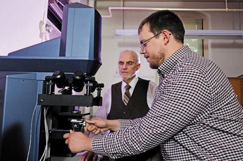
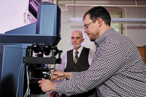
A team at Kansas State University, led by David Wetzel, has been applying this approach to multiple applications, including the analysis of wheat. In particular, the group has worked on alternative methods for the determination of flour and milling stream purity, because outdated methods such as mineral ash residue impurity analysis do not properly reflect the quality of the final products of milling and are dependent upon the soil where the wheat is milled. Mark Boatwright, who is studying for his doctorate under Wetzel, talked to Spectroscopy about some of this work.

You have developed a quantitative near-infrared (NIR) chemical imaging technique to measure endosperm purity in wheat milling process streams, to determine whether the endosperm of a wheat kernel has been fully separated from the surrounding bran layer (1). First, why is such a method needed, and how can such information be useful to flour millers?
Flour milling is a high-volume, low-margin industry. Thus, a method to monitor small differences in product quality and purity is essential to maximize the yield of product. Quantitative NIR chemical imaging provides an alternative to traditional methods that rely on measurement of either the inorganic impurities remaining after ignition of the organic product or the off-color added by residual bran impurities present with the endosperm.
Why did you select NIR as the technique to apply to this problem? And what additional information is provided by using imaging?
NIR spectroscopic imaging provides the depth of penetration necessary for the analysis of coarse, heterogeneous mixtures and is advantageous for quantitative chemical analysis. The use of the imaging technique reveals localized chemical heterogeneity for the mixture.
This approach also allows easy acquisition of large spectral libraries. The technology was originally developed for use in the pharmaceutical industry to study chemicals with discrete spectra. However, a large spectral library is necessary to assess the small chemical differences that are present between various botanical parts of the wheat kernel.
How was statistical analysis used in this study?
To simplify the analysis, the wheat kernel was defined as a binary system, endosperm versus nonendosperm. Spectral libraries in excess of 200,000 individual spectra were defined for each of the two components.
Partial least squares analysis was applied to each sample that was imaged and resulted in chemical identification of each pixel from 1 (endosperm) to 0 (nonendosperm). The summation of the 81,920 pixels in the field of view provided the overall quantitative result.
What were the biggest challenges in developing this method?
Our major challenge has been developing meaningful spectral libraries with the proper spectral window that are compatible for all samples present within the milling process, while reducing the time necessary for data acquisition.
What were you able to achieve with the method?
This method has provided the chemically based contrast required to make operational decisions based on product purity specifications. It also allows millers to monitor the efficiency of the milling operation in terms of the optimum product available from a particular wheat grist, whereas traditionally the measurement of impurity was limited by mineral variability dependent on the soil composition where the wheat was grown.
What are your next steps in this work?
Our next step is to transfer the chemically based technology that we have developed to instruments with potential similar to that of our own and to make it readily available to the milling industry.
The continued goal will be to take advantage of emerging optical technology and to minimize the spectral window to further reduce cost for an industry that has not yet applied quantitative chemical imaging analyses.
Reference
- M.D. Boatwright, E.S. Posner, R. Lopes, and D.L. Wetzel, Cereal Foods Worldwide60(5), 211–216 (2015).


Newsletter
Get essential updates on the latest spectroscopy technologies, regulatory standards, and best practices—subscribe today to Spectroscopy.


Scientists Use Water and Light to Uncover Honey Adulteration
July 30th 2025In a 2025 study, Indian researchers demonstrated that combining near-infrared (NIR) spectroscopy with aquaphotomics enables rapid, non-destructive detection of adulterants in honey by analyzing changes in water’s spectral behavior. Using chemometric models, they accurately identified and quantified six common adulterants, offering a powerful tool for food authenticity and quality control.


Scientists Use AI and Spectroscopy to Detect Fake Honey in Bangladesh
July 29th 2025Researchers in Bangladesh have developed a rapid, non-destructive method to detect honey adulteration using UV-Vis-NIR spectroscopy paired with machine learning. Their findings could protect consumers and support food quality enforcement.


