
|Articles|August 2, 2019
- Special Issues-08-02-2019
- Volume 34
- Issue 8
Fast Microparticle Analysis with Raman: Five Steps to Characterization and Quantification
Author(s)WITec
Advertisement
Microparticle analysis techniques are rapidly evolving in response to demands for regulatory oversight, especially with regard to microplastic pollution. Certain steps are common to any effort to analyze microparticles; They need to be found, classified, individually investigated, identified, and quantified. An automated combination of confocal white-light microscopy and Raman spectroscopy is the most promising approach for these measurements. The following guide describes using such an instrument, the WITec alpha300 R confocal Raman microscope equipped with ParticleScout, to characterize a particulate sample.
1. Choose a sample containing Raman-active microparticles and carry out a large-area survey.
A confocal white-light microscope can use bright- or dark-field illumination to see particles in contrast to the substrate or filter on which they're located. If the area of interest is larger than the microscope objective's field of view, image stitching can automatically combine many areas into a single image. Particles of widely varying height can be accommodated by focus stacking, which acquires images at several focal distances and compiles them to ensure that all particles show sharply defined edges for recognition by software.
2. Select criteria to categorize particles and guide the Raman measurement.
The WITec ParticleScout advanced analysis tool allows located particles to be categorized and ranked in a list by physical properties such as area, perimeter, minimum or maximum Feret diameter, elongation, and many others. The outlines of the selected particles are then used to create a mask that guides the subsequent measurement while omitting all other particles and the empty space in between, which drastically reduces measurement time.
3. Examine the specified category of particles at the individual level.
Raman spectroscopy can detect photons whose energies are shifted slightly from the excitation wavelength to serve as a unique "fingerprint" for each chemical component. The method is nondestructive and doesn't require specialized sample preparation.
Large-area bright-field (A) and dark-field (B) views of a mixture of microplastic particles generated by image stitching. The particles are displayed as a two-color mask (C). From every particle, a single Raman spectrum is acquired (E-F). The chemical compositions of the individual particles are then identified.
The mask created in the previous step guides the excitation light to the particles of interest so the microscope can gather the shifted light and acquire a Raman spectrum from each one. TThis information is stored with the location and category of each particle for further analysis.
4. Chemically identify each particle of interest.
The Raman spectra from each particle can be evaluated manually or using advanced software. For researchers looking for a particular material, the spectra, with their distinctive peaks and contours, will provide all the information required to identify the particles. When the composition of a sample isn't as well established or there are many materials of interest, an integrated Raman database managment software such as WITec's TrueMatch can help expedite identification.
5. Compile the identities of each particle in a table that provides a quantitative sample overview.
With particles chemically characterized, the results can be consolidated and described quantitatively in the form of a table or histogram. This summary shows the researcher exactly what is in their sample, broken down into categories that they selected. It is the detailed and comprehensive answer to the question, "What does this sample contain, and in what proportions?"
Articles in this issue
almost 7 years ago
Vol 34 No 8 Spectroscopy, August 2019, The Resource Issue PDFalmost 7 years ago
Seven Common Errors to Avoid in LIBS Analysisalmost 7 years ago
Seven Essential Steps for In Situ Reaction Monitoringalmost 7 years ago
Key Errors to Avoid in the Consideration of Fluorescence Quenching Dataalmost 7 years ago
A Short Guide for Raman Spectroscopy of Eukaryotic Cellsalmost 7 years ago
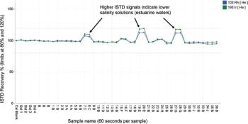
ICP-MS: Essential Steps to Optimize Matrix Tolerancealmost 7 years ago
Spectroscopy Software/Computer Hardware/Automation Productsalmost 7 years ago
Spectroscopic Instrumentation: Spectrometer Systemsalmost 7 years ago
Sampling/Sample HandlingAdvertisement
Related Content
Advertisement

Special Issues
Advertisement
Advertisement
Trending on Spectroscopy Online
1
Where is Raman Spectroscopy Delivering the Most Value for Real-Time Chemical Analysis in Oil and Gas?
2
The Raman Issue 2026
3
Particle Correlated Raman Spectroscopy (PCRS): A Workflow for Correlating Particle Morphology with Chemical Identification
4
Fujifilm, Horiba Unveil New Inline Raman System
5