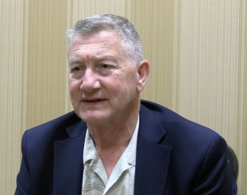
The Versatility of Photothermal MIRSI
Key Takeaways
- Rohith Reddy develops advanced spectroscopic and optical imaging methods for high-resolution cancer detection in tissues like breast, prostate, kidney, and bone marrow.
- His research integrates engineering, computational analysis, and chemometrics to enhance diagnostic accuracy using FT-IR imaging and photothermal mid-infrared spectroscopy.
In this interview segment with Rohith Reddy, he discusses how mid-infrared spectroscopic imaging (MIRSI) can be used to help detect numerous disease types.
Rohith Reddy, associate professor at the University of Houston and CPRIT Scholar in Cancer Research, develops advanced label-free spectroscopic and optical imaging methods for tissue diagnostics (1,2). His group employs Fourier transform infrared (FT-IR) imaging, photothermal mid-infrared spectroscopy, and optical coherence tomography to achieve high-resolution cancer detection in tissues such as breast, prostate, kidney, and bone marrow (1). Trained in engineering and bioengineering, Reddy integrates instrumentation, computational analysis, and chemometrics to enhance diagnostic accuracy (1). A recipient of multiple spectroscopy awards, he presented at SciX 2025 on how optical photothermal infrared (O-PTIR) and mid-infrared frequency combs improve resolution and throughput in diagnosing kidney and bone marrow disorders (2).
Spectroscopy sat down with Reddy to discuss his research more in depth. Our first interview segment concentrated on how Reddy and his team are using IR imaging and photothermal mid-infrared (MIR) spectroscopy to improve tissue analysis (3). In the second part of our conversation with Reddy, he discusses how mid-infrared spectroscopic imaging (MIRSI) can be used to help detect numerous disease types.
Will Wetzel: You’ve demonstrated applications ranging from lupus nephritis to ovarian cancer and bone pathology. What do these diverse case studies reveal about the versatility of photothermal MIRSI as a diagnostic platform across different disease types?
Rohith Reddy: Photothermal MIRSI is relatively new technology compared to FT-IR or other well-known technologies, and it's like it has several advantages compared to false discovery rate (FDR), and the spectroscopic information you get at very high resolution allows us to see things which you previously difficult to see, and allows us to deal with samples which were difficult to analyze before. And so, in my laboratory, we are looking at diagnostics and clinical ovarian cancer, cervical cancer, endometrial cancer, kidney lupus, nephritis, Alzheimer's, like bone marrow fibrosis. We are looking at a wide variety of diseases. The combination of machine learning with the photothermal imaging is, I think, will be revolutionary in terms of, basically, we're using spectroscopy to solve biomedical and clinical problems. I think that's a very big, wide field, and it's very, very promising.
This interview clip is the final part of our interview with Reddy. To stay up to date with the latest coverage of the 2025 SciX Conference, click
References
- University of Houston, Dr. Rohith Reddy. UH.edu. Available at:
https://www.ece.uh.edu/faculty/reddy (accessed 2025-10-13). - SciX Conference, Enhanced Photothermal Mid-Infrared Spectroscopic Imaging for Diagnosing Kidney and Bone Marrow Disorders. SciX Conference. Available at:
https://www.scixconference.org/2025-final-program (accessed 2025-10-13). - Wetzel, W. Improving Resolution and Throughput in Tissue Analysis. Spectroscopy. Available at:
https://www.spectroscopyonline.com/view/improving-resolution-and-throughput-in-tissue-analysis (accessed 2025-10-15).
Newsletter
Get essential updates on the latest spectroscopy technologies, regulatory standards, and best practices—subscribe today to Spectroscopy.


