Article
Application Notebook
Application Notebook
Chemical Analysis of Microscopic Fluorescent Materials by Dispersive 1064 Raman System
Author(s):
Raman measurement on microscopic inclusions in fluorescent materials requires the ability to measure in small volumes, excellent throughput, and long wavelength excitation such as 1064 nm for fluorescence reduction.
Raman measurement on microscopic inclusions in fluorescent materials requires the ability to measure in small volumes, excellent throughput, and long wavelength excitation such as 1064 nm for fluorescence reduction. Carotenoids and keratin can be measured in epoxy and fossil amber by BaySpec's dispersive 1064 confocal Raman microscope.
Inclusions, small particles less than 1 mm in diameter embedded in other materials, are of interest in a number of different areas. In some manufactured products they result in cosmetic, structural, or chemical resistance defects. On the other hand, entrapped inclusions in paleology, or paints and stains in artefacts are a time-capsule from our past. This article demonstrates a few examples of fast, nondestructive identification of microscopic fluorescent samples using Raman spectroscopy, which were previously unattainable.
The challenge for Raman spectroscopy in the past was that it was susceptible to fluorescence. Working with 1064 nm wavelength lasers avoids fluorescence, by avoiding exciting the electronic transitions. Traditionally, due to grating and detector limitations, 1064 nm Raman is often obtained by an FT-Raman system, which is very slow because each spectrum is composed by constantly moving interferometers. Sample mapping using an FT-Raman system is almost impossible. With recent advances in gratings and InGaAs detectors, volume mapping, depth and surface profiling can be accomplished easily with BaySpec's Raman system. For instance, a 1064 nm Raman microscope in dispersive configuration with f2 high-throughput Raman spectrometer is able to create a chemical image as fast as a traditional Raman microscope with visible lasers such as 532 nm. Furthermore, such dispersive configuration allows great flexibility of the system, such as a high-magnification video probe being added to the system to measure the area of interest quickly on samples that do not fit under a microscope.


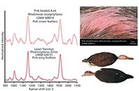
Figure 1: 1064 nm Raman spectra of feathers from an extinct pink-headed duck and from a flamingo wing. Courtesy of Dr. Daniel Thomas, Smithsonian Institution. Refer to reference 1 for Raman spectra of feathers in fossil amber. Raman Data were taken by BaySpec’s Nomadic⢠1064 nm Raman microscope.
Microscopic feathers can occasionally be found embedded in fossil amber, which hold clues about the prehistoric animals. The technique was successfully applied to fossil feathers by using BaySpec's Nomadic™ 1064 nm Raman microscope (Figure 1) (1). The versatility of the video probe in being able to make measurements of areas and depth of a painting is unmatched (Figure 2). In a quick summary, Raman imaging using 1064 nm excitation provides a unique and rapid ability to chemically profile inclusions with fluorescence.


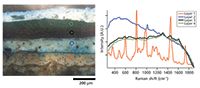
Figure 2: Painting’s cross-section and spectra of the different layers using a Raman video microprobe attached to a BaySpec RamSpec⢠1064 nm Raman spectrometer.
Reference
(1) D.B. Thomas, P.C. Nascimbene, C.J. Dove, D.A. Grimaldi, and H.F. James, Sci. Rep. 4, 5226 (2014).
BaySpec, Inc.
1101 McKay Drive, San Jose, CA 95131
tel. (408) 512-5928
Website: www.bayspec.com


Newsletter
Get essential updates on the latest spectroscopy technologies, regulatory standards, and best practices—subscribe today to Spectroscopy.


