
Spectroscopy Interviews
Latest News
Advertisement
Latest Videos


Shorts
0:50
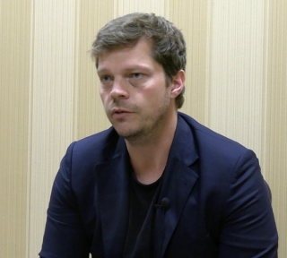
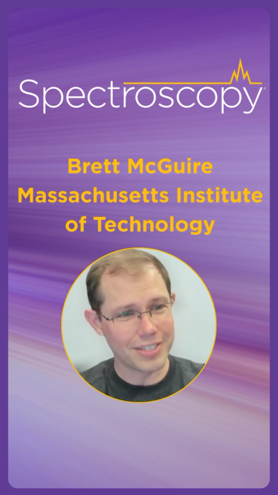
Studying Single and Double Ring Aromatic Molecules
5 months ago
by
Brett McGuire
0:37
The Hidden Enemy in Scientific Analysis: Contamination
5 months ago
by
Anika Retzmann
0:24
How AI Should Be Used in Science
5 months ago
by
Alex Scheeline
0:46
The Benefit of Laminography
5 months ago
by
Johanna Nelson Weker
2:47
Pathways in Spectroscopy: How to Be A Successful Product Manager
5 months ago
by
John Margeson
1:20
Is SIBS Just Repeating the History of LIBS?
5 months ago
by
Alex Scheeline

0:59
The Ongoing Trends and Developments in Imaging Spectroscopy
5 months ago
by
Christian W. Huck
1:38
Conducting Faster Elemental Mapping Using GDOES
5 months ago
by
Gerardo Gamez
0:51
The Remaining Challenges with Field Deployability
5 months ago
by
Tom Metz
1:14
Data Quality and Reproducibility When Analyzing Zinc Isotope Composition in Mice Organs
5 months ago
by
Anika Retzmann
Advertisement
More News

Spectroscopy recently sat down with Dr. Geraldine Monjardez and two of her coauthors, Dr. Christopher Zall and Dr. Jared Estevanes, to discuss their most recent study, which examined the crystal structure of ammonium nitrate (AN) following exposure to explosive events.
In part 2 of our interview with Oskar Hagelskjaer of Microplastic Solution, he discusses the benefits of using automated Raman microspectroscopy to detect and analyze microplastics in drinking water.
Spectroscopy sat down with Oskar Hagelskjaer, Founder and CEO of Microplastic Solution, to discuss his latest study whose findings challenge EU Directive 2020/2184 regarding microplastic detection in potable water.
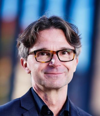
Spectroscopy sat down with Juergen Popp of the Leibniz Institute for Photonic Technology to talk about the Photonics West Conference, as well as his work using label-free spectroscopy techniques for precise tumor margin control.
Spectroscopy sat down with Knut Baumann of the University of Technology Braunschweig to discuss his latest research examining the classification of two closely related horsetail species, Equisetum arvense (field horsetail) and Equisetum palustre (marsh horsetail), using near-infrared spectroscopy (NIR).
Spectroscopy sat down with Daniel Cozzolino of the University of Queensland to discuss his latest research using near-infrared (NIR) spectroscopy to determine the fatty acid content in black soldier fly.
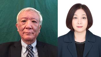
Spectroscopy recently sat down with Isao Noda of the University of Delaware and Young Mee Jung of Kangwon National University to talk about the principles of two-dimensional correlation spectroscopy (2D-COS) and its key applications.
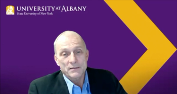
During EAS 2024, we interviewed Igor Lednev of the University of Albany about his storied career and his expectations for 2025.

Aleksandra "Sasha" Karapetrova and Win Cowger discuss their research using µ-FTIR spectroscopy and Open Specy software to investigate microplastic deposits in remote snow areas, shedding light on the long-range transport of microplastics.

Spectroscopy spoke with Uwe Karst, a full professor at the University of Münster in the Institute of Inorganic and Analytical Chemistry, to discuss his research on hyphenated analytical techniques in battery research.
As part of “The Future of Forensic Analysis” content series, Spectroscopy sat down with Glen P. Jackson of West Virginia University to talk about the historical development of mass spectrometry in forensic analysis.
As part of “The Future of Forensic Analysis” content series, Spectroscopy sat down with Brooke Kammrath of the University of New Haven to talk about the significance of spectroscopy in forensic analysis.
As part of our EAS 2024 coverage, we recently interviewed Rachel Martin of the University of California, Irvine about her work and her being awarded the EAS Award for Outstanding Achievements in Magnetic Resonance.
Spectroscopy sat down with Landulfo Silveira Jr. of Universidade Anhembi Morumbi-UAM and Center for Innovation, Technology and Education-CITÉ (São Paulo, Brazil) to talk about his team’s latest research using Raman spectroscopy to detect biomarkers of cancer in canine sera.
As part of our SciX 2024 conference coverage, we recently asked Anita Mahadevan-Jansen of Vanderbilt University about how mentorship has impacted her career and how new scientists can find a mentor.
As part of our SciX 2024 conference coverage, we recently asked Ellen Miseo of Miseo Consulting about how mentorship has impacted her career and how new scientists can find a mentor.
Spectroscopy Magazine sits down with Benjamin T. Manard of the Chemical Sciences Division at Oak Ridge National Laboratory (Oak Ridge, Tennessee), who will be receiving the Lester W. Strock Award, given by the New England Section of the Society for Applied Spectroscopy, this October.
In a preview to the upcoming SciX Conference October 20 to 25 in Raleigh, North Carolina, Spectroscopy sat down with Nick Stone of the University of Exeter to discuss his recent work in oncology and clinical analysis.
In this preview interview for SciX 2024, we talk with Conor Evans of Harvard Medical School about his research with sparse spectral sampling stimulated Raman scattering (S4RS) and his excitement for the upcoming conference.
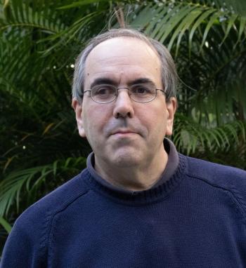
In this preview interview for SciX 2024, Jason Dwyer of the University of Rhode Island discusses his experience with SERS and his feelings on winning the American Electrophoresis Society's Mid-Career Award.

Matthieu Baudelet, an associate professor of Chemistry at the National Center for Forensic Science at the University of Central Florida, is currently exploring how laser-based spectroscopic techniques can be used in forensic anthropology. Spectroscopy recently sat down with Matthieu Baudelet, Kristen Livingston, and Katie Zejdlik to discuss their research as part of “The Future of Forensic Analysis” content series.
As part of “The Future of Forensic Analysis” content series presented by Spectroscopy, we sat down with Dr. Rajinder Singh of Department of Forensic Science, Punjabi University, Patiala, to talk about his recent work using attenuated total reflectance Fourier transform infrared spectroscopy (ATR-FT-IR) to distinguish different animal species based on hair samples.
As part of "The Future of Forensic Analysis," executive editor Jerome Workman, Jr. sat down with Igor Lednev to discuss several of his recent papers related to his spectroscopic research in forensic analysis.

A joint French-Canadian study examined the ripening process of commercially popular Comté and cheddar cheeses, which are widely consumed in those countries, utilizing mid-infrared (mid-IR) and synchronous fluorescence spectroscopy (SFS) in their analysis.

A recent article authored by scientists from the Institute of Sport and Preventive Medicine, part of the University of Saarland (Saarbrücken, Germany), discusses their investigation of the absolute and relative test-retest reliability of the Moxy Monitor, as well as their investigations into side differences of oxygen saturation at the vastus lateralis muscle of both legs in male cyclists.
Advertisement
Advertisement
Trending on Spectroscopy Online
1
When Political Appointees Outrank Peer Review: How a New Office of Management and Budget Rule Could Reshape Spectroscopy and Scientific Research Funding
2
The State of Spectroscopy Careers
3
Is Produced Water a Source of Lithium?
4
The Importance of Hands-On Learning
5