Spectroscopy Interviews
Latest News

Harishchandra Singh, Graham King and associates have employed high energy synchrotron X-ray diffraction (HE-SXRD) experiments and an analytical model in order to predict the yield strength of cerium-modified super duplex stainless steel (SDSS) subjected to various cold- and cryo-deformation. Spectroscopy recently had the opportunity to discuss the experiments and the findings with Singh and King.
Raman Spectroscopy and Machine Learning-Based Optical Probe for Tuberculosis Diagnosis via Sputum

Quantitative Mapping of Mercury and Selenium in Mushroom Fruit Bodies with Laser Ablation–Inductively Coupled Plasma-Mass Spectrometry
Latest Videos

More News

Wei Min, of the Department of Chemistry at Columbia University in New York City, and his associates recently published a paper outlining their devising a set of multiplexed Raman molecular probes with sharp and mutually resolvable Raman peaks to simultaneously quantify cell surface proteins, endocytosis activities, and metabolic dynamics of an individual live cell. Min, who recently spoke to us about this work, is the 2022 recipient of the Craver Award, presented annually at FACSS SciX to recognize the efforts of young professional spectroscopists that have made significant contributions in applied analytical vibrational spectroscopy.
Igor Gornushkin and colleagues at BAM Federal Institute for Materials Research and Testing in Berlin, Germany studied the feasibility of using optical spectroscopy as a control method for laser metal deposition, and he recently spoke to us about this work. Gornushkin is the 2022 recipient of the Lester W. Strock Award from the New England Chapter of the Society for Applied Spectroscopy (SAS).
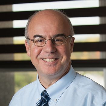
Noureddine Melikechi of the Department of Physics and Applied Physics at the University of Massachusetts (Lowell, MA) saw an urgent need for the development of an untargeted and unbiased method to distinguish Gulf War illness (GWI) patients from non-GWI patients; he and his associates utilized laser-induced breakdown spectroscopy (LIBS) in their efforts to meet that need.

Spray paint is often used by vandals for creating graffiti, as well as for criminals to leave signs, messages, and blots to conceal the left traces at the scene of their efforts. Rajinder Singh and his colleagues in the Department of Forensic Science at Punjabi University (Punjab, India) have used attenuated total reflectance Fourier transform infrared (ATR-FT-IR) spectroscopy for nondestructive analysis of 20 red spray paints of different manufacturers, which could possibly be encountered at a crime scene, particularly in case of vandalism. Singh spoke to Spectroscopy about the findings, and the paper that resulted from their efforts.

Mohamed Abdel-Harith of Cairo University and his team explored using BRELIBS for the elemental analysis of the synagogue’s glass shards. Their findings reveal the potential of BRELIBS in conducting elemental analysis on transparent materials. Spectroscopy recently spoke to Abdel-Harith about this work.
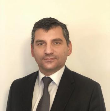
Meat fraud has emerged as a growing concern globally. The adulteration, substitution, or mislabeling of meat products has resulted in negative economic effects, such as unfair competition in the global marketplace, as well as ethical concerns, because consumers want to know what is in the meat they are consuming. Ismail H. Boyaci of Hacettepe University and his team have explored using laser-induced breakdown spectroscopy (LIBS) for protein-based analysis and the identification of meat species. Spectroscopy recently spoke to Boyaci about this work.
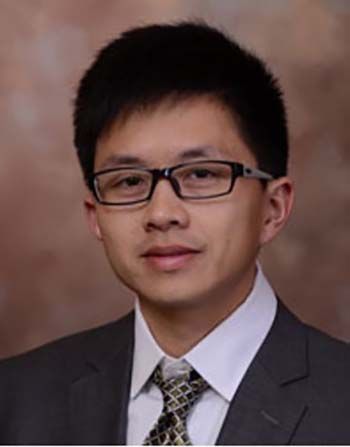
Determining the quality of the food we consume is important not just for reasons of safety, but for verifying authenticity as well. Changmou Xu, a Research Associate Professor at the University of Nebraska-Lincoln (UNL), and his colleagues have been exploring methods for food analysis that are rapid but do not harm the environment or the analysts.
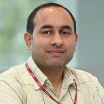
As global food supplies and security have been challenged by water scarcity and climate variations, the expected increase in food demand will require a corresponding increase in crop productivity and disruptive improvements in agricultural production systems, including implementing strategies to mitigate the degradation of crop yield caused by plant diseases. Several groups have explored the use of Raman spectroscopy for rapid diagnosis of such diseases.

Denise M. Mitrano is an Assistant Professor of Environmental Chemistry of Anthropogenic Materials at ETH Zurich in the Department of Environmental Systems Science. Her research is directed to understanding the impact and interaction of nanoparticles in the environment using atomic spectroscopy techniques, such as inductively coupled plasma–mass spectrometry (ICP-MS) and single-particle ICP-MS (sp-ICP-MS). She is the winner of the 2022 Emerging Leader in Atomic Spectroscopy Award. Chosen by an independent committee, the Emerging Leader in Atomic Spectroscopy Award recognizes the achievements and aspirations of a talented young atomic spectroscopist who has made strides early in his or her career toward the advancement of atomic spectroscopy techniques and applications. We recently interviewed Mitrano about her work.

As forensic analysis continues to advance, such as in the understanding of source identification and analysis of trace quantities of bodily fluids, spectroscopic techniques and machine learning are playing a significant role. Igor K. Lednev, a chemistry professor at the University at Albany, SUNY, in Albany, New York, has been working in this field with his team. The analytical methods currently under investigation include Raman spectroscopy, attenuated total reflection Fourier transform infrared (ATR FT-IR) spectroscopy, and advanced chemometric classification and analysis methods. We recently interviewed him about his work.

A tutorial and spreadsheet for the validation and bottom-up uncertainty evaluation of quantifications performed by instrumental methods of analysis based on linear weighted calibrations were presented by Ricardo J.N. Bettencourt da Silva of the University of Lisbon in Lisbon, Portugal, and colleagues. This software tool was successfully applied to the determination of the mass concentration of Cd, Pb, As, Hg, Co, V, and Ni in a nasal spray by ICP-MS after samples dilution and acidification. Bettencourt da Silva spoke to Spectroscopy about applying this software tool and the implications for a better understanding of quantitative analytical results.
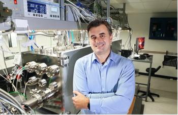
The limited availability of table-top extreme ultraviolet (XUV) sources with adequate fluxes and coherence properties has resulted in a lack of nonlinear XUV and X-ray spectroscopies to free-election lasers (FELs). Michael Zuerch of the Department of Chemistry at the University of California, Berkeley, along with colleagues from ten laboratories across three countries, has conducted the first extreme ultraviolet second harmonic generation (XUV-SHG) experiment above the Ti M edge (32.6 eV), also representing the first table-top demonstration of SHG at photon energies beyond the UV regime.

Emmanuel Lalla of York University in Toronto, Ontario, Canada, as well as his co-authors, recently presented an in-depth characterization of a set of samples collected during a 28-day Mars analog mission conducted by the Austrian Space Forum in the Dhofar region of Oman. Lalla spoke to Spectroscopy about this research, as well as how various methods of spectroscopic analysis can complement each other in analysis.
Katsumasa Fujita, a professor of applied physics at Osaka University, has been working to improve techniques for imaging biological samples using spontaneous Raman scattering.
Salvatore La Cavera III and his colleagues in the Faculty of Engineering at the University of Nottingham, in Nottingham, United Kingdom have developed a novel measurement system consisting of two ultrafast lasers that excite and detect high-frequency ultrasound from a nano-transducer fabricated onto the tip of a single-mode optical fiber.
Zac Schultz of The Ohio State University and his team used tip-enhanced Raman spectroscopy (TERS) and surface-enhanced Raman spectroscopy (SERS) with gold nanostars to investigate chemical reactions involved in protein–ligand binding. He recently spoke with Spectroscopy about his findings.

Bhavya Sharma is the winner of the 2021 Emerging Leader in Molecular Spectroscopy Award. We recently interviewed her about her work conducting research to detect active and important biomolecules related to hormone regulation, neurological health, and disease diagnosis.
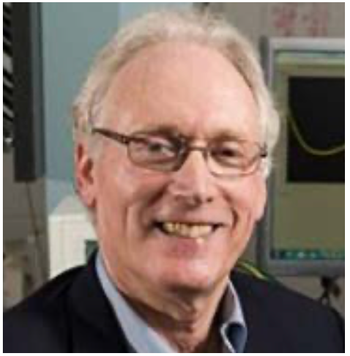
Mark Meyerhoff has been exploring chemical sensors for biomedical applications. Because of his work, Meyerhoff has been awarded the 2021 ANACHEM award. Meyerhoff spoke to us about his work, his career, and what being presented this award at this fall’s SciX event means to him.
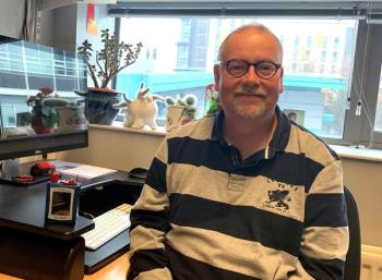
Roy Goodacre, a professor of biological chemistry at the University of Liverpool in the United Kingdom, first used SERS to achieve whole-organism fingerprinting of bacteria and then explored SERS in a variety of other applications, including within biotechnology, disease diagnostics, quantitative detection, imaging, food security, and more. Goodacre is the 2021 winner of the Charles Mann Award for Applied Raman Spectroscopy. This interview is part of an ongoing series of interviews with the winners of awards that are presented at the annual SciX conference.
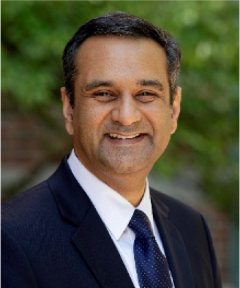
Professor Rohit Bhargava and his team at the University of Illinois, where they have established the Cancer Center at Illinois, are advancing research in tumor microenvironments, using techniques such as high-definition Fourier transform infrared (HD-FT-IR) coupled with machine learning. We recently spoke to Bhargava about this work.
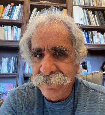
Analytical chemists are continually striving to advance techniques to make it possible to observe and measure matter and processes at smaller and smaller scales. Professor Vartkess Ara Apkarian and his team at the University of California, Irvine have made a significant breakthrough in this quest: They have recorded the Raman spectrum of a single azobenzene thiol molecule. The approach, which breaks common tenets about surface-enhanced Raman scattering/spectroscopy (SERS) and tip-enhanced Raman spectroscopy (TERS), involved imaging an isolated azobenzene thiol molecule on an atomically flat gold surface, then picking it up and recording its Raman spectrum using an electrochemically etched silver tip, in an ultrahigh vacuum cryogenic scanning tunneling microscope. For the resulting paper detailing the effort [1], Apkarian and his associates are the 2021 recipients of the William F. Meggers Award, given annually by the Society for Applied Spectroscopy to the authors of the outstanding paper appearing in the journal Applied Spectroscopy. We spoke to Apkarian about this research, and what being awarded this honor means to him and his team. This interview is part of an ongoing series with the winners of awards that are presented at the annual SciX conference. The award will be presented to Apkarian at this fall’s event, which will be held in person in Providence, Rhode Island, September 28–October 1.
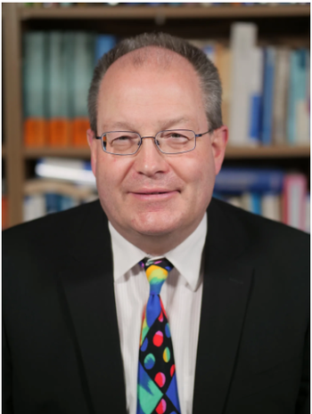
Uwe Karst of the University of Münster in Germany explains the use of laser ablation–inductively coupled plasma–mass spectrometry (LA-ICP-MS) imaging to provide spatially resolved quantification of trace elements in biological samples.
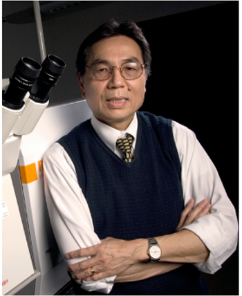
Working at the frontiers of biotechnology, fiberoptics, lasers technique, and molecular spectroscopy, Tuan Vo-Dinh of Duke University has developed multiple sensor technologies for medical research and diagnostics. Throughout this work, Vo-Dinh and his research colleagues have brought spectroscopy to biomedical applications. In this second recent interview, Vo-Dinh talks about his research work and philosophy.
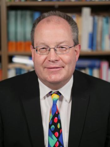
Uwe Karst of the University of Münster in Germany explains the use of laser ablation–inductively coupled plasma–mass spectrometry (LA-ICP-MS) imaging to provide spatially resolved quantification of trace elements in biological samples.
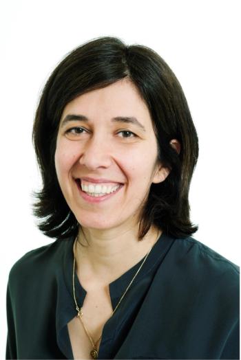
Metallomics approaches based on mass spectrometry have become increasingly important in the support of developing metal-based anticancer drugs. This area is a key focus for Gunda Koellensperger and her colleagues at the University of Vienna (Austria) and they recently published an article discussing this state-of-the-art instrumentation, as well as highlighting recent analytical advances, focusing especially on the latest developments in inductively coupled plasma-–mass spectrometry (ICP-MS).

