Spectroscopy Interviews
Latest News
August 20th 2024
By John Chasse
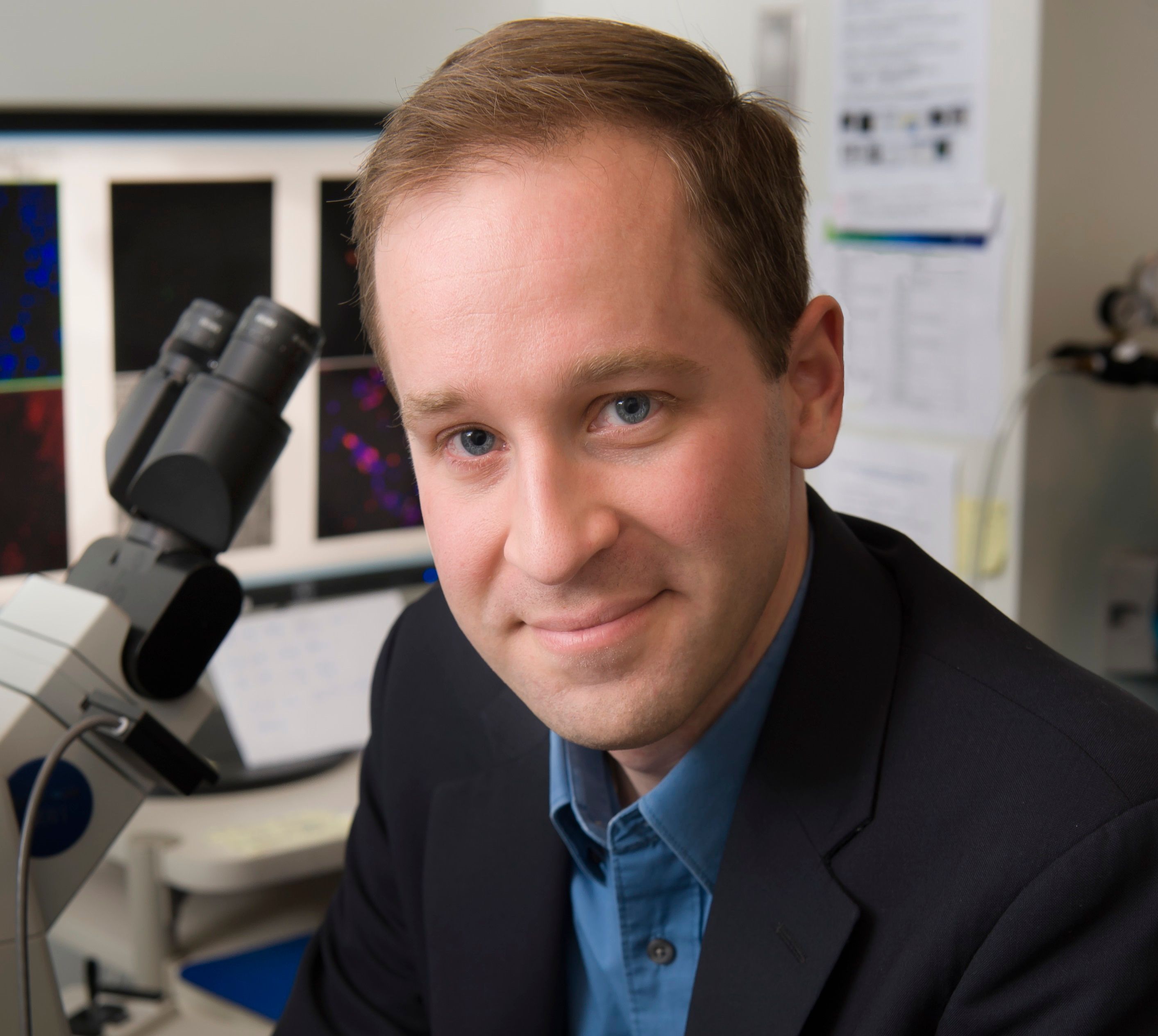
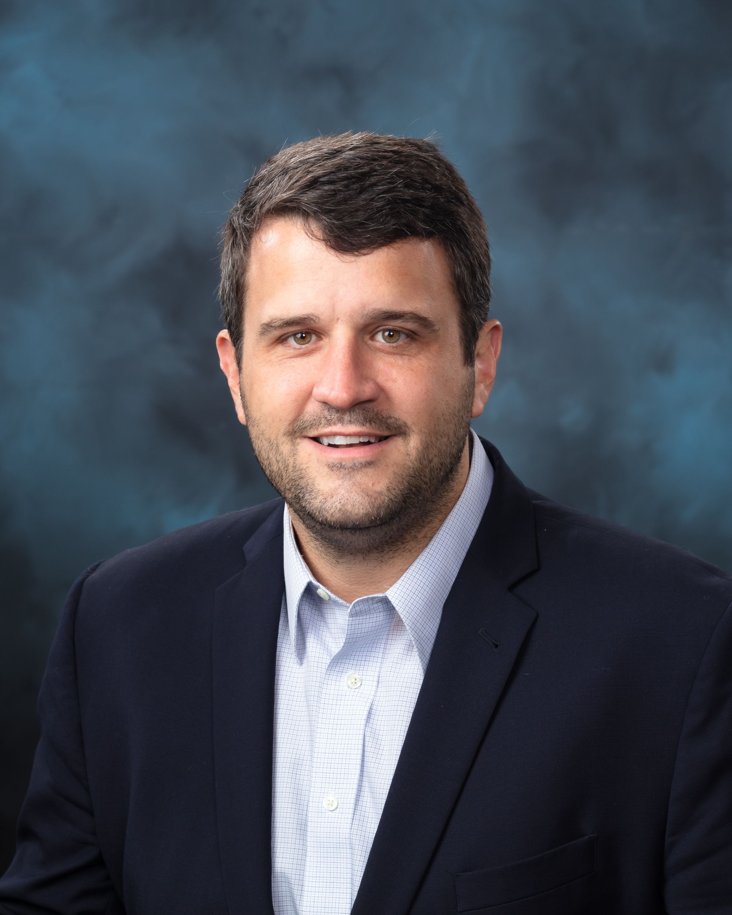
Spectroscopy Magazine sits down with Benjamin T. Manard of the Chemical Sciences Division at Oak Ridge National Laboratory (Oak Ridge, Tennessee), who will be receiving the Lester W. Strock Award, given by the New England Section of the Society for Applied Spectroscopy, this October.
Advertisement
Latest Videos
Shorts
1:14
Data Quality and Reproducibility When Analyzing Zinc Isotope Composition in Mice Organs
5 months ago
by
Anika Retzmann
1:56
What Strategic Decisions Have the Greatest Long-Term Impact on Businesses?
5 months ago
by
Mercedes Bertotto
2:27
The Surprising Realities Spectroscopists Face in Business
5 months ago
by
Mercedes Bertotto
0:59
How a Humanitarian DNA Database is Solving Cold Cases Across Borders
5 months ago
by
Claire Glynn
0:48
How Particle Size Affects Local Electric Fields
5 months ago
by
Nishadi Nadeeshani Moragoda Liyanage
4:17
What Skills Do Entrepreneurs Have to Learn Quickly After Starting Their Business?
5 months ago
by
Mercedes Bertotto
0:47
The Biggest Mistake Scientists Make: Not Using the Right Technique
5 months ago
by
Johanna Nelson Weker
0:38
Using Optical Spectroscopy to Monitor Nuclear Radiation Safely
5 months ago
by
Hunter Andrews
2:52
How Working in a National Reference Laboratory Led to a New Opportunity Overseas
5 months ago
by
Mercedes Bertotto
0:52
How Compressed Sensing Is Revolutionizing Data Acquisition
5 months ago
by
Gerardo Gamez
Advertisement
More News
Uwe Karst of the University of Münster took time at Analytica 2024 to talk about the session he's chairing, as well as detail the future of imaging and other trends in analytical chemistry.
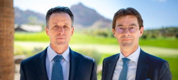
Spectroscopy spoke with the CEO and CCO of the instrument manufacturer PerkinElmer to discuss trends in the analytical and life sciences industry.

Dulasiri Amarasiriwardena, emeritus professor of chemistry at Hampshire College, Amherst, Massachusetts, and his team have been conducting research using laser ablation-inductively coupled plasma-mass spectrometry (LA-ICP-MS) to investigate trace metal nutrition and exposure to toxic metal(loid) pollutants by studying Andean mummy remains. We sat down with Prof. Amarasiriwardena to discuss his research.
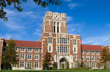
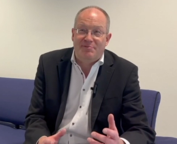
Tessa Calhoun, PhD, an associate professor of biochemistry & cellular and molecular biology and chemistry at the University of Tennessee – Knoxville, discusses her group’s most recent work employing the optical technique, second harmonic scattering (SHS), to probe how living bacterial membranes uptake and transport small molecules, including antibiotics.
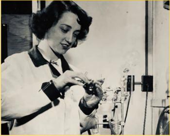
As one of the first renowned women spectroscopists, Craver broke down barriers in the field and is recognized for her work compiling reference libraries of spectra that set the foundation for modern infrared (IR) spectroscopy.
Narangerel Altangerel, Zhenhuan Yi, and Marlan Scully of Texas A&M University recently used TRIP to analyze eight protein–ligand systems. Spectroscopy recently spoke to these three researchers about their findings and what the implications are for high-throughput drug screening.
At SPIE Photonics West, Spectroscopy spoke with Ana Doblas of the University of Massachusetts, Dartmouth about artificial intelligence, deep learning, and their potential in microscopy.
LCGC and Spectroscopy Editor Patrick Lavery spoke with Oligo Factory CEO Chris Boggess about the company’s recently attained compliance with Good Manufacturing Practice (GMP) International Conference on Harmonisation of Technical Requirements for Registration of Pharmaceuticals for Human Use (ICH) Expert Working Group (Q7) guidance and its distinction from Research Use Only (RUO) and International Organization for Standardization (ISO) 13485 designations.

At the Winter Conference on Plasma Spectrochemistry, Jacob Shelley of Rensselaer Polytechnic Institute sat down with Spectroscopy to talk about the latest work he and his group are conducting.

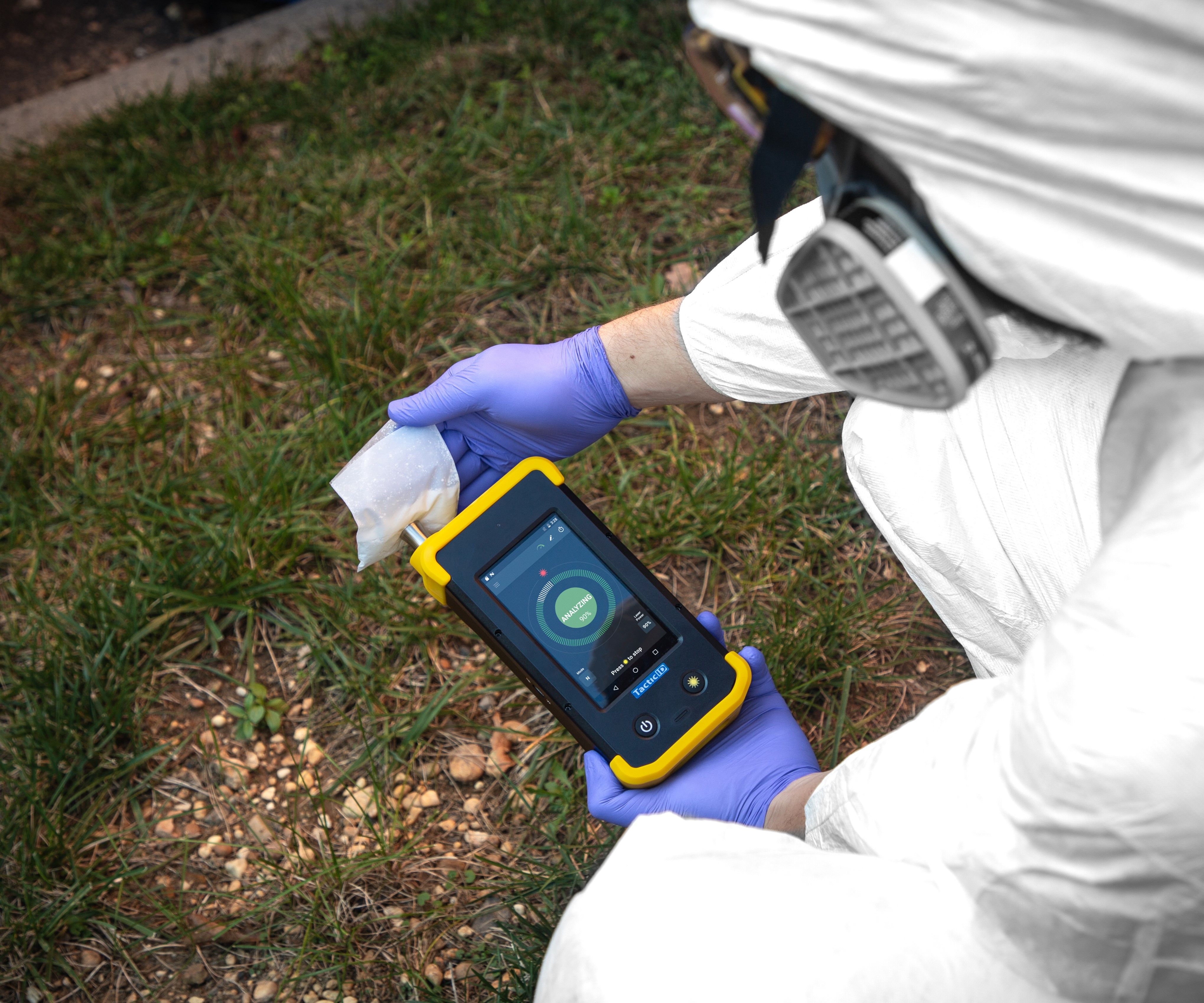
Miri Park of the Fraunhofer Institute for Environmental, Safety, and Energy Technologies is examining how Raman spectroscopy could aid non-destructive sensing in agricultural science. Recently, Park sat down with Spectroscopy to discuss micro-Raman spectroscopy's role in assessing crop quality, particularly secondary metabolites, across different contexts (in vitro, in vivo, and in situ), while suggesting future research for broader application possibilities.
In this edition of “Inside the Laboratory,” John Cottle, PhD, a professor of geology at the University of California, Santa Barbara, and a member of Spectroscopy’s Editorial Advisory Board, discusses his group’s most recent work using “laser ablation split steam” analysis to measure elemental concentrations and isotopic ratios in rocks and minerals.
At the Winter Conference on Plasma Spectrochemistry, John Burgener of Burgener Research spoke with us about his company's nebulizers and detailed his proudest achievement.
At the Winter Conference on Plasma Spectrochemistry, Spectroscopy sat down with Robert Jones to discuss how he used ICP-MS to advance the work of the Center for Disease Control (CDC).
At the Winter Conference on Plasma Spectrochemistry, Spectroscopy magazine sat down with John Burgener of Burgener Research Inc. to discuss his career in inductively coupled plasma (ICP) and the importance of accumulating various experiences during your career.

Spectroscopy spoke with researchers from the Columbia Climate School about how they are using stimulated Raman scattering microscopy to test for nanoplastics in water bottles.
At the Winter Conference on Plasma Spectrochemistry, Spectroscopy sat down with Robert Jones to discuss his career at the Center for Disease Control (CDC), and how their ICP-MS laboratory helped advance the work of the CDC.
At the Winter Conference on Plasma Spectrochemistry, Spectroscopy magazine sat down with John Burgener of Burgener Research Inc. to discuss his career working with mass spectrometers, inductively coupled plasma (ICP), and developing the Burgener nebulizer.

The Surface-Specificity of IRRAS in Studying Soluble Organic Acids: An Interview with Alexandra Deal
Spectroscopy spoke with Alexandra Deal about her latest research in using infrared reflection absorption spectroscopy (IRRAS) to analyze molecules at the surface level.
At the Eastern Analytical Symposium (EAS) in Plainsboro, New Jersey, Spectroscopy sat down with Robert Kennedy to discuss his research and career in analytical chemistry.
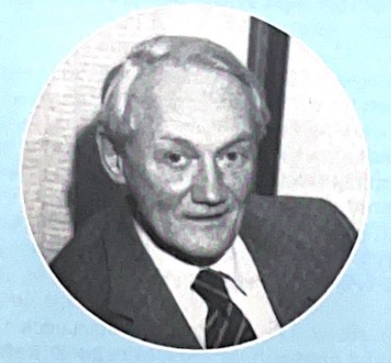
At the Eastern Analytical Symposium (EAS) in Plainsboro, New Jersey, Spectroscopy sat down with Curtis Marcott to discuss his research and career in spectroscopy.

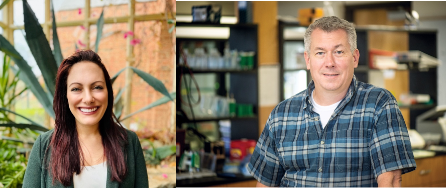
Monica Arienzo, an associate research professor in the Division of Hydrologic Sciences at the Desert Research Institute, and her team recently used ATR-FT-IR to determine the polymer composition of plastic litter recovered by scuba divers from the lakebed of Lake Tahoe. Spectroscopy spoke to Arienzo about the significance of her work and how spectroscopy can be used to help monitor plastic litter in the environment.
At the 2023 Gulf Coast Conference, Spectroscopy spoke with Kevin Schug of the University of Texas at Arlington, about predicting gas phase vacuum ultraviolet spectra using machine learning. This interview was one of four conducted live at GCC 2023.
At the 2023 Gulf Coast Conference, Spectroscopy spoke with Elena Hagemann of Metrohm USA, about spectroscopy in relation to petrochemicals. This interview was one of four conducted live at GCC 2023.
Spectroscopy sat down with 2023 Lester W. Strock Awardee Maria Montes-Bayon to talk about her research and what winning the Strock award means to her.
Fresh off his plenary talk at SciX, Wei Xiong sat down with Spectroscopy magazine to talk about his career accomplishments and what he enjoys most about his job.
Advertisement
Advertisement