Sponsored Content
See how Raman microscopy in combination with SEM, AFM, topographic imaging, and other methods can characterize properties of geoscience samples.
Sponsored Content
See how Raman microscopy in combination with SEM, AFM, topographic imaging, and other methods can characterize properties of geoscience samples.

Webcasts
Webinar Date/Time: Thu, Sep 14, 2023 10:00 AM EDT
Sponsored Content
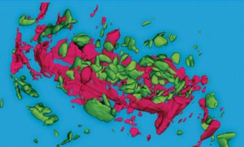
This application note shows correlative Raman SEM-EDS imaging investigations of Li-ion batteries that reveal charge cycle-induced chemical and structural changes.
Sponsored Content
See how Raman microscopy in combination with SEM, AFM, topographic imaging, and other methods can characterize chemical and structural properties of geoscience samples.
Sponsored Content
This application note shows correlative Raman-SEM-EDS imaging investigations of Li-ion batteries that reveal charge cycle-induced chemical and structural changes.
Sponsored Content
This white paper describes applications of cryoRaman, a cryogenic Raman microscope offering VIS to NIR excitation, high magnetic fields, and full polarization control.
Sponsored Content
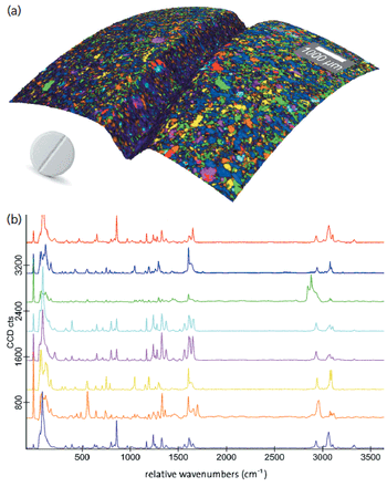
This overview of advanced microparticle analysis shows how particles can be found, classified, and then identified automatically using confocal Raman microscopy.

This application note describes a correlative Raman–scanning electron microscope (SEM) investigation of a lithium-ion battery cathode before and after hundreds of charge cycles. The technique provides an exceptionally detailed chemical and structural analysis of degradation processes on the nanoscale.
Sponsored Content
This survey shows how Raman imaging can characterize food samples such as honey, chocolate, and fat spreads to help understand the products and production processes.
Sponsored Content
This white paper describes applications of cryoRaman, a cryogenic Raman microscope offering VIS to NIR excitation, high magnetic fields, and full polarization control.

Sponsored Content
This study describes the generation of 3D images of pharmaceutical, biological, and geological samples using image stacking enabled by a highly confocal beam path.
Sponsored Content
This overview of advanced microparticle analysis shows how particles can be found, classified, and then identified automatically using confocal Raman microscopy.
Sponsored Content
This compilation provides an introduction to the principles of Raman imaging and shows examples of correlative variations including Raman–SEM, Raman–AFM, and others.
Sponsored Content
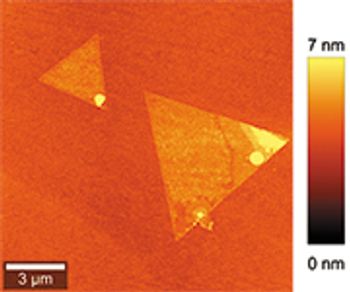
This study of molybdenum disulfide illustrates the advantages of Raman, second harmonic generation (SHG) and photoluminescence (PL) microscopy for investigating TMDs.
Sponsored Content
This survey shows how Raman imaging can characterize food samples such as honey, chocolate, and fat spreads to help understand the products and production processes.
Sponsored Content
This overview of advanced microparticle analysis shows how particles can be found, classified, and identified automatically using confocal Raman microscopy.
This study shows Raman imaging, atomic force microscopy, and scanning near-field optical microscopy characterizing and depth-profiling polymer blends and layers.
See how Raman microscopy in combination with SEM, AFM, topographic imaging, and other methods can characterize chemical and structural properties of geoscience samples.

This study describes how Raman imaging and correlative Raman scanning electron (RISE) microscopy can characterize and visualize layer numbers in 2D MoS2 and WSe2 samples.

The quality and stress field properties of gallium nitride (GaN) crystals must be investigated in order to refine production processes. 3D Confocal Raman microscopy is uniquely capable of the chemical c and visualizing strain in this material.
Sponsored Content
This study shows Raman imaging, atomic force microscopy and scanning near-field optical microscopy characterizing and depth-profiling polymer blends and layers.
Sponsored Content
WITec Application Note: ParticleScout for Automated Confocal Raman Imaging Analysis of Microparticles

High-resolution measurements of particles are of great interest in many fields of application. With ParticleScout, WITec has developed a tool that makes it possible to find, classify, and identify particles automatically.
Sponsored Content
This overview of advanced microparticle analysis shows how particles can be found, classified and then identified automatically using confocal Raman microscopy.

Special Issues
This application note describes the advantages of a simplified process of searching through Raman spectral databases and creating new ones for example, based on user generated data.
Application Notebook
Application Notebook
Application Notebook

September 1st 2019
March 8th 2023

February 1st 2022

September 1st 2021
