Peer-reviewed Articles

Latest News

Dotlich-fig11_web.jpg
February 1st 2017

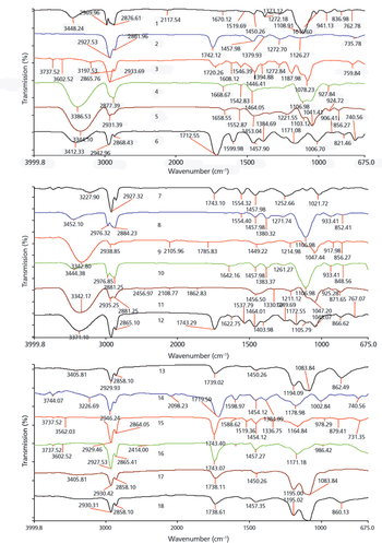
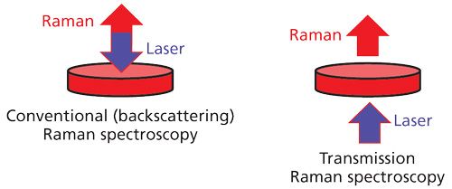
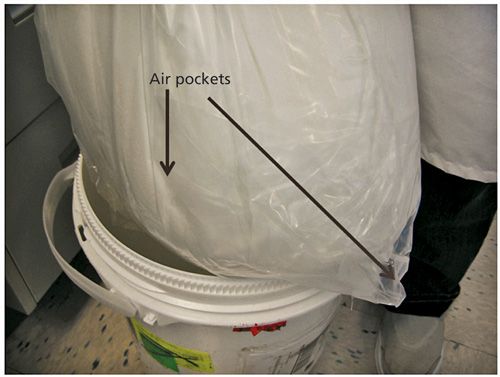
Portable instrumentation for Raman spectroscopy has rapidly evolved over the last decade, where sample testing that once occurred in the laboratory is now executed in the field (e.g. warehouse).   Portable Raman spectroscopy is a powerful technique for the rapid identification of diversely sourced raw materials used in pharmaceutical processing.  In addition to portability; reduced cost, rapid data acquisition and ease of use make this powerful technique attractive and accessible to both expert spectroscopists and non-specialists.  In most cases, the method development can be easily accomplished in the laboratory after which the instrument and methods are transferred to field for sample analysis or warehouse areas for inspection of incoming raw material.  Qualitative Raman methods for identification of raw materials typically utilize spectral libraries for sample to standard comparison.  When developing Raman spectral libraries for raw material identification, great care is required when considering critical factors (e.g. instrument type, Raman capability, container type, container interference, background interference, material variability) that can potentially influence the identity of the material.  This paper discusses portable Raman techniques and approaches for raw material identification, as well as key considerations for developing and validating Raman spectral libraries.


Bhargava-fig01-web.jpg

Spectroscopy

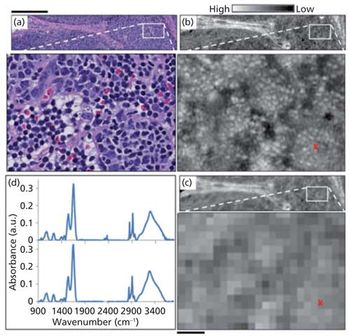
Recent advances in instrumentation have enabled new forms of vibrational chemical imaging, including discrete frequency infrared (DFIR) microscopy and stimulated Raman scattering (SRS) microscopy. These technologies may represent a fundamental shift in how we approach spectroscopic imaging: rather than collecting full spectra which contain redundant information, measuring a few important spectral frequencies may enable significant gains in speed, throughput, signal to noise ratio, and/or image quality. For infrared microscopy, these advantages may be compounded by High Definition IR microscopy. Here we discuss recent advances in infrared and nonlinear Raman imaging through the lens of 'discrete frequency' approaches, including several examples of applications and critical issues in instrumentation that are likely to be dominating research themes in the near future.

table1-web-3.jpg

Spectroscopy

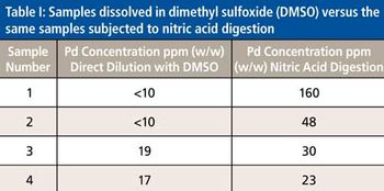
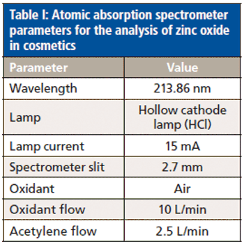
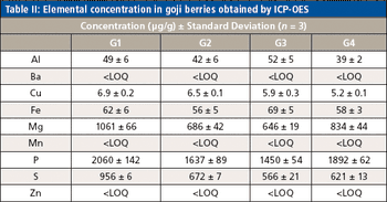
The use of atomic spectroscopy techniques and sample preparation procedures is something that is not as routine in the pharmaceutical industry as are chromatography-based techniques and sample preparation procedures. With new requirements being implemented regarding elemental impurities by the United States Pharmacopoeia (USP) and International Conference on Harmonization (ICH), analysts in the pharmaceutical industry are, in many cases, working to determine how best to analyze their samples. Sample preparation techniques that can be used for pharmaceutical samples are the same ones that have been used by other industries for many years. This paper will provide a brief overview of potential techniques.

Gordon-figure04_web.jpg

Spectroscopy

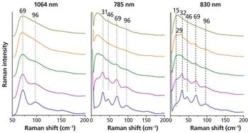
Crystallinity is an important factor when producing pharmaceuticals as it directly affects the bioavailability of the drug. Low frequency Raman spectroscopy offers some advantages to the detection and analysis of crystallinity in pharmaceutical samples. Here the experimental requirements for low frequency Raman measurements are described. The application to the study of crystallinity with a number of examples is discussed and the advantages and limitations of this technique are highlighted and compared with other techniques.

Wu-figure1_web.jpg

Spectroscopy

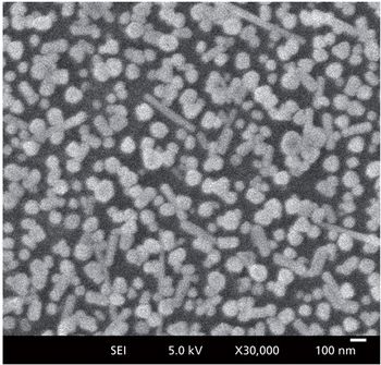
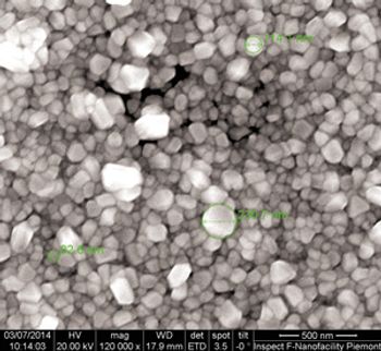
In this paper, we demonstrate a sensitive surface-enhanced Raman spectroscopy (SERS) substrate for trace dimethoate detection. The substrate is composed of Ag nanoparticle/probe/smooth Au film stack configuration. The nanogap formed by an Ag nanoparticle and a macroscopically flat Au film is one kind of “hot site” which will dramatically increase the total “hot spots” number.

Myrick-figure_2_WEB.jpg

Spectroscopy

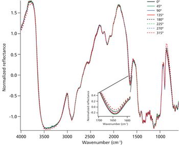
Infrared spectroscopy is an appealing technique for application to forensic samples because it offers the benefits of being non-destructive and non-hazardous, fast, reasonably sensitive, and resistant to some of the interferences of many commonly used techniques. Our research team has been focusing on detecting biological fluids on fabrics, which are inherently anisotropic substrates for spectroscopy. The work presented here investigates the effect of azimuthal angle of the sample on the infrared diffuse reflection spectra of fabrics with a goal of removing sampling differences as a source of analytic variation.

Figure-6b_web.jpg

Spectroscopy

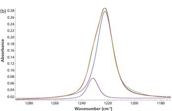
Infrared and Raman bands are typically described in terms of peak location and peak height. The shape of the peak-its form, width, and asymmetry-is less well understood. Bandshapes largely depend upon interactions between vibrating molecules and their environment. An understanding of this relationship may enhance spectral interpretation and can explain unexpected behaviors, in both qualitative and quantitative analyses. This paper presents basic concepts that influence line shapes and shows the complex lineshape changes in an apparently simple system of acetone and water.

Spec1015_White_fig07_web.jpg

Spectroscopy

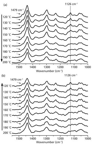
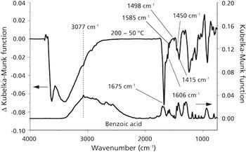
High stability Fourier transform-infrared (FT-IR) interferometers make it possible to conduct experiments designed to identify subtle sample structure changes resulting from external perturbations. In particular, IR spectrum measurements obtained while heating samples can be used to associate specific structure changes with incremental additions of thermal energy. This paper describes the application of variable temperature diffuse reflection infrared Fourier transform spectroscopy (VT-DRIFTS) for analysis of benzoic acid adsorbed on montmorillonite clay. The technique described here is generally applicable for soil adsorbate characterization studies, and therefore should be useful for elucidating soil transport mechanisms for selected contaminants.

Tucker-figure01_web.jpg

Spectroscopy

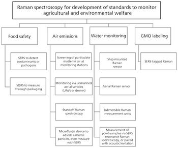
Raman spectroscopy is applied to quality control of agricultural products with greater frequency, and can also be used to refine regulatory criteria for both agricultural and environmental monitoring. It is now integrated into everything from hand held SERS detectors to unmanned aerial vehicles to monitor the gamut from genetic variation to soil and water content. Development opportunities, particularly with bundled technologies, continue to emerge as demand for quality assurance increases.

Salafranca_fig02_web.jpg

Spectroscopy

Surface-enhanced Raman scattering (SERS) has been applied to the determination of the antioxidant butylated hydroxyanisole (BHA), commonly used in fatty foods and oils to prevent their oxidation. The use of SERS-Raman microscopy with an inexpensive homemade silver substrate allowed the direct determination of BHA in oils without any sample handling. Several edible and essential oils (used as flavorings) have been considered for this purpose.

Spectroscopy3_i26.jpg

Spectroscopy

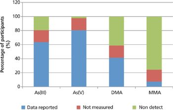
An interlaboratory comparison study for the measurement of arsenic species in rice, kelp, and apple juice was carried out. The purpose of the study was to enable participating laboratories to evaluate their analytical capability to determine inorganic arsenic, arsenite, arsenate, monomethylarsonic acid, and dimethylarsinic acid, assess the intercomparability of the data generated, and look for any correlation trends between the results and the analytical procedures used.

Spectroscopy5_i6-859589-1417772811319.jpg

Spectroscopy

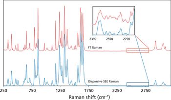
A method for removing fluorescence-induced backgrounds from Raman spectra using sequentially shifted excitation (SSE) is described. The method uses a temperature-tuned, distributed Bragg reflector diode laser to produce the multiple excitation spectra required to implement the SSE algorithm. Examples applying the SSE method to analysis of motor oils and edible oils are given.

Spectroscopy4_i4_t-848105-1416903144204.jpg

Spectroscopy

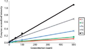
Determination of trace metals in oil and petroleum typically is based on ICP-OES methods, requiring signficant sample preparation and expensive instrumentation. This article presents an alternative method, based on the use of a handheld energy-dispersive X-ray fluorescence analyzer, that involves minimal sample preparation, uses authentic standards for calibration, gives low parts-per-million detection limits, and provides significant time and cost savings.