


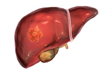
Scientists recently tested a new means of detecting early-stage liver cancer using surface-enhance Raman spectroscopy (SERS).
Scientists recently tested a new means of detecting early-stage liver cancer using surface-enhance Raman spectroscopy (SERS).
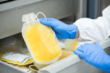
In a new study, scientists have created a fluorescence chemo sensor to selectively determine histamine in human plasma samples.

A group of Brazilian scientists tested a new means of testing for COVID-19 using a combination of Vis-NIR spectroscopy and machine learning algorithms.
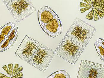
Using single-cell inductively coupled plasma mass spectrometry (SC-ICP-MS), scientists from Kanazawa, Japan created a new method for detecting cadmium in marine phytoplankton.

Ishan Barman, the 2023 recipient of the Coblentz Society Clara Craver Award, held a plenary session during SciX that focuses on transformative biophotonics in disease detection and monitoring.

The Coblentz Society created the Clara Craver Award to recognize young individuals who have made significant contributions in applied analytical vibrational spectroscopy. The work may include any aspect of infrared (IR), terahertz (THz), or Raman spectroscopy in applied analytical vibrational spectroscopy. This year’s recipient, Ishan Barman, is an Associate Professor in the Department of Mechanical Engineering at the Johns Hopkins University with joint appointments in Oncology and Radiology and Radiological Science.

To effectively classify tobacco stems and impurities, a group of scientists from Jiangsu, China used hyperspectral superpixels to separate classify compounds and avoid the influence of interference fringes.

A group of scientists from the University of Wrocław and the University of Innsbruck studied the relationships between aliphatic ketones and water molecules in various environments.

Scientists from the Amirkabir University of Technology in Tehran, Iran, used laser-induced fluorescence (LIF) spectroscopy to analyze opium and hashish, in order to achieve a novel method of drug analysis.

A team of scientists created a portable infrared attenuated total reflection (IR-ATR) spectrometer and tested its food analysis capabilities against similar systems.

In a recent study, scientists used a combination of SERS, butanol, and gold nanoparticles to analyze the virus.
Scientists used laser induced breakdown spectroscopy (LIBS) to analyze the safety of food.

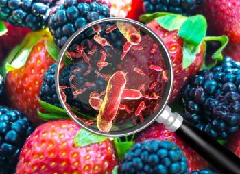
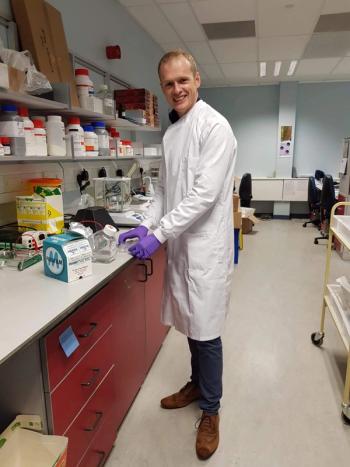
In this interview, James Chapman discusses his current and future research efforts, and how combining spectroscopy with machine learning tools can change how bacterial research is conducted.
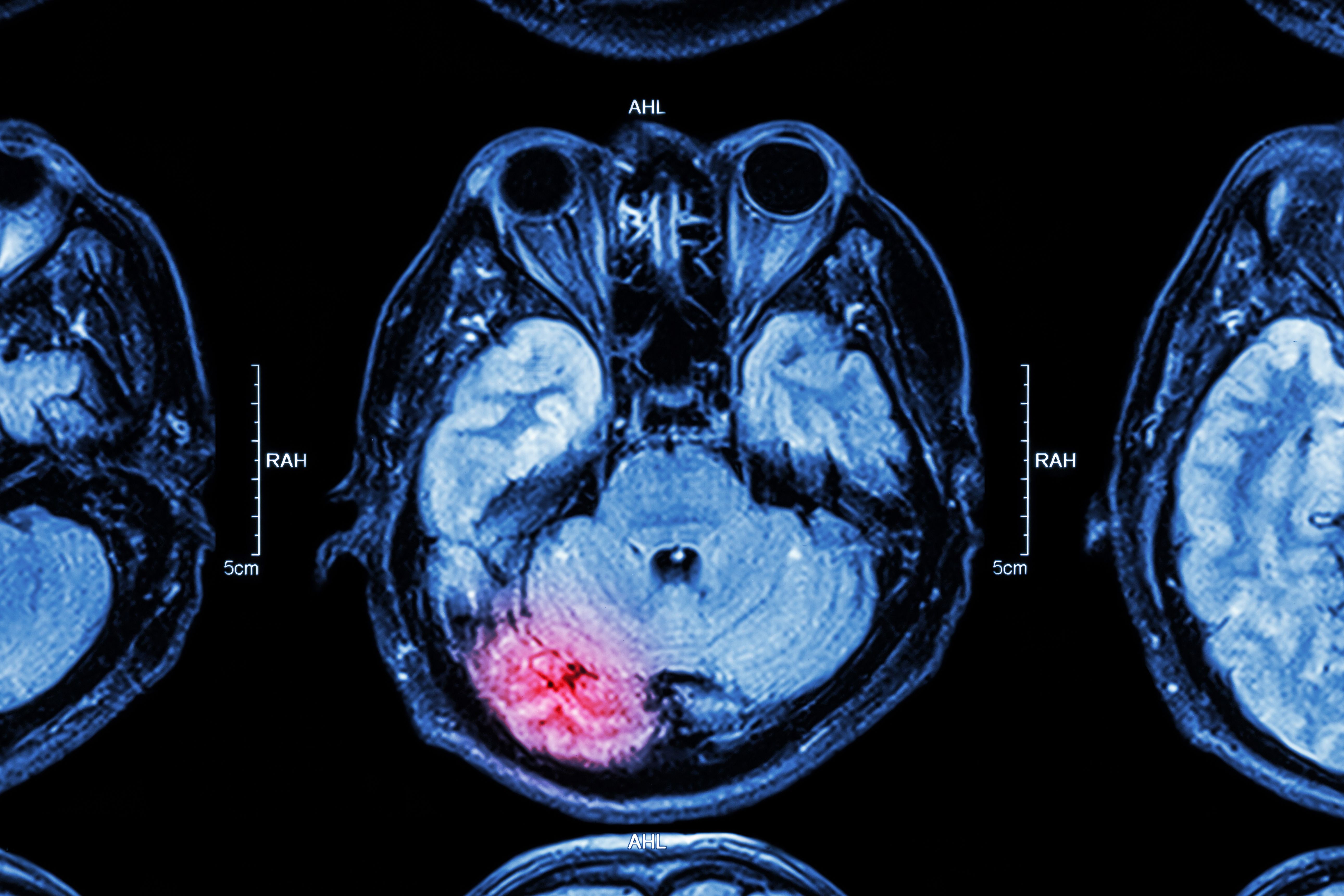
Mathew Horrocks, the 2023 recipient of The Joseph Black Prize, shares his thoughts about his current work developing and using single-molecule and super-resolution microscopy techniques to study amyloid oligomers and their commonality regarding a variety of neurodegenerative disorders.

In this interview, Jürgen Popp discusses the importance of Raman spectroscopy, where it can make a difference, and how it can be evolved and improved on in the future.

A team of researchers has conducted a successful round-robin test using total reflection X-ray fluorescence (TXRF) to analyze the elemental composition of rat tissue samples. The preliminary results demonstrate the effectiveness of TXRF in accurately determining the elemental composition of mammalian tissue.

Researchers have developed a near-infrared fluorescent probe that allows for highly sensitive detection of butyrylcholinesterase activity and pesticide residue in food samples. The probe offers a ratiometric pattern and shows promise for applications in health evaluation, disease diagnosis, and environmental monitoring.

A research team has utilized the Allan variance technique to analyze the performance characteristics of compact Fourier transform infrared (FT-IR) spectrometers. The study provides insights into the noise sources and instabilities of these handheld instruments, offering guidance for improving their accuracy and stability in real-time material detection and quantification applications.

Scientists have conducted a spectroscopic analysis of 2-amino-1-naphthalenesulfonic acid, exploring its electronic properties and its potential as an antiviral agent.

Biomedical Raman imaging is growing in the biomedical space, where technical advances and new information processing tools and techniques aim to propel the field into the future. An upcoming conference in Atlanta, Georgia, will explore these developments while bringing scientists in this field together.
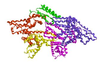
A recent study explored the binding characteristics of the tyrosine kinase inhibitor entrectinib with bovine serum albumin through multi-spectral analysis and theoretical calculations, revealing the factors affecting the stability of the ENB-BSA complex and potential ways to enhance the efficacy of ENB.

This interview with Young Jong Lee highlights the work he and his team have done to reinvent solvent absorption compensation (SAC), and the potential it has across multiple forms of spectroscopy.

Terahertz time-domain spectroscopy was used to identify and analyze the low-frequency vibrational modes of three free anthraquinones, revealing the vibrational contributions of different atoms and groups.
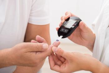
SERS of centrifuged blood serum samples of diabetic type II patients using 50 KDa filter devices can diagnose the disease at an early stage by studying small molecular weight proteins.

Researchers have developed a method for the simultaneous determination of cadmium and lead in water samples using microwave-induced plasma optical emission spectrometry with multiwalled carbon nanotubes pre-concentration and a discrete sample introduction system.