



Surface-enhanced Raman spectroscopy (SERS)
Latest News
Advertisement
Latest Videos



Shorts
0:31
Best of the Week: Implementing SERS in Clinical Settings, How Lasers Became Valuable Tools in Spectroscopy
5 months ago
by
Will Wetzel
Advertisement
More News

Wednesday's program at SciX 2026 kept returning to one question: can spectroscopy perform outside the controlled laboratory? Speakers took surface-enhanced Raman scattering (SERS), laser-induced breakdown spectroscopy (LIBS), mid-infrared photonics, chemometrics, and fluorescence excitation–emission matrix (EEM) spectroscopy into clinics, food plants, reactor coolant loops, brine fields, and drinking water supplies.
Raman spectroscopy is entering one of its most consequential periods since its commercialization, driven by artificial intelligence, coherent heterodyne detection, and radical instrument miniaturization, with new deep-learning frameworks and portable systems moving Raman analysis into fields, factories, and crime scenes.
Raman spectroscopy just got a brain, a stopwatch, and a nose. Together, these upgrades are turning a century-old light-scattering trick into a frontline tool for catching cancer earlier, chiral drugs cleaner, and toxic chemicals faster than ever before.
Reliability, dynamic measurements, artificial intelligence, and real-world applications are reshaping Raman spectroscopy – while Molecular Photonics points toward new translational frontiers.
Raman spectroscopy is shedding its bulky, benchtop reputation as chip-scale spectrometers, tip-enhanced probes, and AI-native detectors push the technique into pockets, production lines, and single molecules. The result is an instrument category being rebuilt from the optics up, faster, smaller, deeper, and smarter than the Raman systems of even five years ago.

Researchers developed spherical and hollow gold nanoparticle-based SERS platforms that enable label-free molecular imaging of chemotherapy drug effects and drug release within cancer cells, offering a theranostic tool with potential applications in early cancer diagnostics.

Measuring neonicotinoids in drinking water is not heavily regulated in the United States, but low-cost, rapid analytical tools are changing the conversation.

A recent study examined how surface-enhanced Raman spectroscopy (SERS) is being used with machine learning (ML) analysis to accurately distinguish patients with Crohn’s disease.
A new diagnostic platform may help detect drug-induced liver injury better. How would this work in real-world clinical settings?
The combination of surface-enhanced Raman spectroscopy (SERS) and digital microfluidics (DMF) might be able to improve detection of liver injury biomarkers.

A recent study showcased a wearable, glove-mounted sensor that can detect trace residues of two cancer drugs on workplace surfaces.

In this interview clip, Sian Sloan-Dennison, a postdoctoral research associate at the University of Strathclyde, discusses how a new digital microfluidics surface-enhanced Raman spectroscopy (DMF-SERS) platform be implemented in clinical settings.
Why is determining the function of a surface-enhanced Raman spectroscopy (SERS) assay important? This "Pathways in Spectroscopy" clip offers career advice for researchers in biological and clinical analysis by addressing this question.
Why does integrating a surface-enhanced Raman scattering (SERS) assay with a digital microfluidics (DMF) platform improve sensitivity, reproducibility, and throughput when detecting onset of drug-induced liver injury?
An upcoming two-part video series on “Pathways in Spectroscopy” tackles working with surface-enhanced Raman spectroscopy (SERS), offering practical advice for researchers in clinical laboratories.
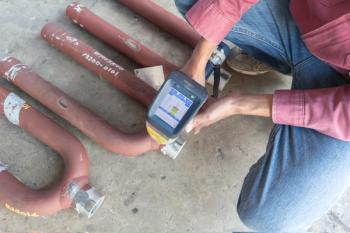
What does the rise of portable and handheld spectroscopic instrumentation tell us?

Top articles published this week include a couple previews of upcoming “Pathways in Spectroscopy” episodes and an inside look at the role spectroscopy is playing in modern agriculture.
In 2025, the vibrational-spectroscopy community saw a convergence of deep learning, advanced simulation, and portable instrumentation that materially changed how spectra are interpreted and applied.
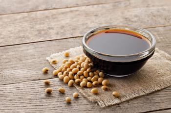
In this study, surface-enhanced Raman spectroscopy (SERS) and solvent extraction were used to detect aconitine (AC) in various complex matrices using gold nanorod substrates. The experimental results demonstrated that ether efficiently extracted AC from soy sauce as an example complex matrix.

Researchers recently developed a nanoscale sensor for rapid detection of GenX in water, enhancing environmental monitoring and safety efforts.

A sweeping review, “Surface-Enhanced Raman Spectroscopy: A Half-Century Historical Perspective” traces the evolution of SERS from its foundational experiments in the 1970s through the nanoscience era and modern high-spatial-resolution techniques.
Top articles published this week include several video interviews from our coverage of the SciX Conference and an inside look at classical correction methods.
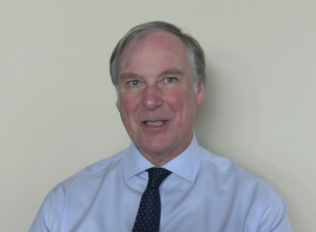
In this segment, Bell addresses the key barriers that are preventing SERS from being more widely adopted in complex environments.
At SciX, Zac Schultz of The Ohio State University sat down with Spectroscopy to discuss his work characterizing molecular interactions with localization microscopy techniques, as well as give his thoughts about how spectrally resolved surface-enhanced Raman spectroscopy (SERS) imaging will impact in vitro cell imaging and other application areas.
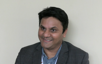
In this interview segment, Prashant Jain of the University of Illinois Urbana-Champaign discusses the unique reaction pathways observed in light-driven chemistry on nanoparticles, particularly in CO2 reduction, and the insights gained into the quantum mechanical mechanisms of these reactions.
Advertisement
Advertisement
Trending on Spectroscopy Online
1
Why Big Data Is Transforming Environmental Nanoparticle Analysis
2
Monitoring Sugar Consumption and Ethanol Production During Yeast Fermentation Using a Raman Plate Reader and Multivariate Analysis
3
Highly Substituted Benzene Rings—The Rest of the Story II: Spectra
4
Best of the Week: Chemometrics Terminology Guide, Pushing Past the Micron Scale
5