


Scientists from Florida International University and Escola Universitària Salesiana de Sarrià Passeig are working on a novel approach to differentiate the structure of fentanyl analogues.
Scientists from Florida International University and Escola Universitària Salesiana de Sarrià Passeig are working on a novel approach to differentiate the structure of fentanyl analogues.

This quarter Spectroscopy spoke to experts in laser ablation, SERS, and Raman spectroscopy, among others.

The authors sought to expand the limitations of Raman spectroscopy applications that can be caused by the fluorescence admitted by some samples.

Here are the top five articles that the editors of Spectroscopy published this week.

Scientists from Duke University in Durham, North Carolina recently tested a surface-enhanced Raman scattering (SERS) spectroscopy system for detecting the SARS-CoV-2 virus.

Pakistani scientists recently used surface-enhanced Raman spectroscopy (SERS) to characterize the metabolites of sulfuric compounds.

In a new study, researchers from Ulsan National Institute of Science and Technology and Pohang University of Science and Technology presented a new surface-enhanced Raman spectroscopy (SERS) device, improving gap plasmon resonance.
Chinese scientists recently made a surface-enhanced Raman scattering (SERS)-based approach to detect synthetic antioxidants in food samples.
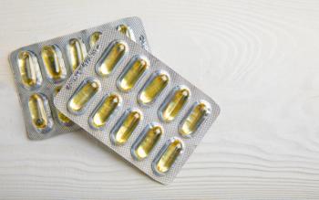
Metrohm has launched a website that details the use of portable Raman spectroscopy instrumentation to detect fentanyl, and other manufacturers are joining the battle too.

A recent study from the University of Shanghai for Science and Technology used surface-enhanced Raman spectroscopy (SERS) to improve analysis of biological fluids such as urine.

In a study led by scientists from Northwest University in Xi’an, China, carbon quantum dots were tested on how well they can detect hypochlorite and aid in cellular imaging.

A recent study from Spain used surface-enhanced Raman spectroscopy (SERS) to study cancer cells with methylthioadenosine phosphorylase (MTAP) deletions, shedding new insights into the metabolic interactions inside the tumor microenvironment that could influence cancer aggression.
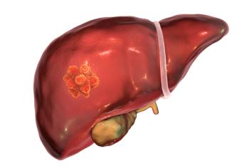
Scientists recently tested a new means of detecting early-stage liver cancer using surface-enhance Raman spectroscopy (SERS).

Duke University researchers, led by Joy Q. Li, revolutionize biomedical diagnostics with a multiplexed SERS-based nanosensor called inverse molecular sentinel (iMS) for micro-RNA detection, employing machine learning, particularly convolutional neural networks (CNN) and non-negative matrix factorization (NMF), to achieve higher accuracy in spectral unmixing, paving the way for more precise and efficient clinical diagnostics.

Researchers at Nanjing University of Science and Technology, led by corresponding have introduced an advancement in surface-enhanced Raman spectroscopy (SERS) with two-dimensional amorphous titanium dioxide/silver (a-TiO2/Ag) nanosheets, demonstrating exceptional sensitivity and repeatability in chemical detection applications.

Ascorbic acid (AA), melatonin (Mel), glutathione (GSH), tea polyphenols (TPP), and uric acid (UA) were distinguished in this experiment by three analyses: heat map, hierarchical cluster, and linear discriminant.

In a recent study, scientists used a combination of SERS, butanol, and gold nanoparticles to analyze the virus.

Researchers have developed a new biosensors decorated with silver nanoparticles that enable the sensitive detection of the Acyclovir drug on filter paper substrates.

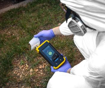
There is a growing desire among spectroscopists for having instruments small enough to be taken to the sample, as opposed to bringing the sample to the instrument. The result is that Raman spectrometers are becoming more miniaturized. Because these instruments come at a lower cost and offer distinct advantages over traditional spectrometers, the expectation is that a rapid expansion of when these instruments are applied will come forthwith. We offer a preview of how future miniaturized Raman spectrometers might look.

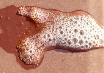
Researchers have created a flexible SERS substrate based on Au nanostars and PDMS, enabling highly sensitive detection of thiram residue in apple juice. The innovative substrate offers a reliable method for ensuring food safety by accurately identifying pesticide contaminants.
Researchers have developed an ultrasensitive and rapid detection method using surface-enhanced Raman spectroscopy (SERS) for multiple dopings in saliva and urine, offering potential advancements in doping control measures.

Researchers explore the impact of sandwich-type DNA construction and plasmonic metal on the signal generated by surface-enhanced Raman scattering (SERS) DNA sensors, giving insight on optimization strategies for improved detection.

A new study has developed a low-cost, sensitive SERS substrate using silver nanoparticle functionalized paper for the detection and analysis of rotavirus in clinical stool samples.

Factor analysis (FA) of the time series of surface-enhanced Raman scattering (SERS) spectra was used to reveal changes in water arrangement and surface plasmon extinction (SPE) in silver nanoparticle systems, which could help to interpret SERS results more accurately.
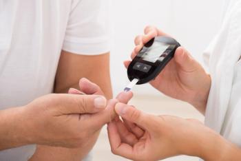
SERS of centrifuged blood serum samples of diabetic type II patients using 50 KDa filter devices can diagnose the disease at an early stage by studying small molecular weight proteins.