


Biopharmaceuticals Biotechnology and Protein Analysis
Latest News
Advertisement
Latest Videos

Shorts

1:00
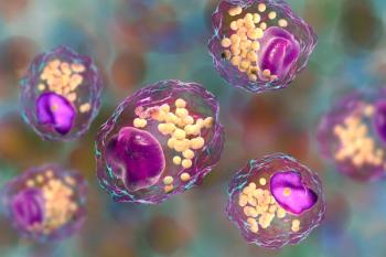
Optimizing Biotherapeutics: Reducing Particle Content for Better Therapies
10 months ago
by
Jerome Workman, Jr.
Advertisement
More News

Researchers from Northern Border University in Saudi Arabia have developed a rhein-loaded self-nano emulsifying drug delivery system (RS-SNEDDS), significantly enhancing the compound's solubility, bioavailability, and therapeutic potential, particularly for neurological applications.

Ludwig-Maximilians-Universität München researchers uncover crucial protein dynamics in lyophilized drugs.

Researchers at Nagoya University and RIKEN have developed a novel computational method to enhance the resolution of high-speed atomic force microscopy (HS-AFM) images for studying protein conformational transitions. The algorithm, normal mode flexible fitting-atomic force microscopy (NMFF-AFM), leverages normal-mode analysis to derive precise molecular models, potentially transforming the understanding of biomolecular dynamics.

Joseph P. Smith, Director of Process R&D Enabling Technologies at Merck has been awarded the 2024 Emerging Leader in Molecular Spectroscopy Award, recognizing his significant contributions to the advancement of molecular spectroscopy in the pharmaceutical industry.

This review highlights very recent advancements in several key spectroscopic methods, including atomic, vibrational, molecular, electronic, and diffraction techniques.

Scientists from the Czech Republic recently created new practices to study different Tröger’s base isomers.

Henan University scientists recently developed a new deep learning-based prediction model for classifying nonclassical secreted proteins.

Howard University scientists recently analyzed tryptophan (Trp) networks and conditions under ultraviolet superradiance.

Scientists from the University of Utretcht in The Netherlands recently created a new autoantibody analysis system based around antigen-binding fragments.

Scientists from Banaras Hindu University in Varanasi, India recently studied how acarbose (ACA) and quercetin (QUE) can help prevent conditions like Type 2 diabetes.

Scientists from Duke University in Durham, North Carolina recently tested a surface-enhanced Raman scattering (SERS) spectroscopy system for detecting the SARS-CoV-2 virus.

Scientists from the Lodz University of Technology in Lodz, Poland created a new system to analyze normal and cancer human colon cells.

Chinese scientists recently created a dual-response fluorescent probe to better understand the pathological mechanisms of depression disorder.

Scientists in Poland recently tested how multidimensional discriminant analysis and reflectance spectroscopy can help identify species within fungi strains.

In a recent study from Ghent University and Charles University, scientists analyzed layered manganese oxide structures using different Raman spectroscopy techniques.

A recent study explores the use of another fluorescent probe that can be used in targeted protein degradation.

In a new study, scientists are investigating Raman spectroscopy as a technique for monitoring postmenopausual osteoporosis.

Scientists from Hirosaki University Graduate School of Science and Technology in Japan evaluated the eating quality of white rice samples using Raman spectroscopy.

High-resolution magnetic angle spinning nuclear magnetic resonance (HRMAS NMR) spectroscopy in particular can be applied to nonconventional solvents, helping to obtain information about protein crystal structures while also adhering to green analytical chemistry tenets.
Scientists from the University of Jinan developed a new fluorescent probe to better understand the relationship between certain lipids.

A team of scientists created a portable infrared attenuated total reflection (IR-ATR) spectrometer and tested its food analysis capabilities against similar systems.
Mathew Horrocks, the 2023 recipient of The Joseph Black Prize, shares his thoughts about his current work developing and using single-molecule and super-resolution microscopy techniques to study amyloid oligomers and their commonality regarding a variety of neurodegenerative disorders.

Researchers have developed a non-destructive method for identifying monoclonal antibody drug substances using Raman spectroscopy.

This interview with Young Jong Lee highlights the work he and his team have done to reinvent solvent absorption compensation (SAC), and the potential it has across multiple forms of spectroscopy.

Raman spectra can help determine protein structure. Here’s how.
Advertisement
Advertisement
Trending on Spectroscopy Online
1
Expansion of a Greener Method of Sex Determination from Hair Using Electrothermal Vaporization Coupled to Inductively Coupled Plasma–Optical Emission Spectrometry
2
UV–FT-IR Studies on α-Lactalbumin-Infused Nickel Hydroxide Nanoparticles
3
From Single-Technique Analysis to Multimodal Characterization: Recent Advances and Future Perspectives for Carbon Nanotubes and Graphene
4
What is the Gordon Research Conference on Vibrational Spectroscopy?
5