


A recent study from Cork, Ireland, used x-ray fluorescence to study three Irish chalices to learn more about Irish history and art.
A recent study from Cork, Ireland, used x-ray fluorescence to study three Irish chalices to learn more about Irish history and art.

Spectroscopic analytical techniques are crucial for the analysis of environmental samples. This review emphasizes the latest advancements in several key spectroscopic methods, including atomic, vibrational, molecular, electronic, and X-ray techniques. The applications of these analytical methods in detecting contaminants and other environmental applications are thoroughly discussed.

A recent study discusses the recent upgrades to the infrared (IR) beamline at BESSY II storage ring, which has helped it improve its results when conducting IR microscopy.

Here are the top five articles that the editors of Spectroscopy published this week.
In a study led by scientists from Northwest University in Xi’an, China, carbon quantum dots were tested on how well they can detect hypochlorite and aid in cellular imaging.

A PhD student in the Department of Bioengineering at the University of Pennsylvania has won the 2024 Physics of Medical Imaging Student Paper Award, which is given out annually by the International Society for Optics and Photonics (SPIE), at the Medical Imaging Symposium in San Diego, California.
Triethylaminium picrate (TEAP) crystals were developed using a slow evaporation solution growth method and then characterized for their feasibility in optoelectronic and other uses.
Studying historical ancient artifacts requires the use of a nondestructive technique to analyze the metal surfaces of these objects. This study presents two approaches that improves on existing methods when conducting alloy analysis.

Researchers at Kochi University and RIKEN have unveiled a new method for distinguishing individual polyester fibers in forensic investigations. Published in Spectrochimica Acta Part B: Atomic Spectroscopy, their advanced X-ray analysis refreshes how we unravel the composition of these fibers.
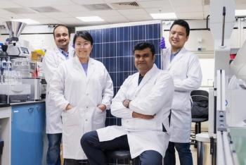
The team devised a method for recovering high-purity silicon from expired solar panels to produce lithium-ion batteries for electric cars.

Scientists faced challenges when using XRF on artifacts that contain many layers.
In a new study, a group of researchers explored the performance of multi-hole collimators with varying geometries and material compositions in a full-field XRF spectrometer based on the 2D-THCOBRA gas detector.

A new study published in Spectrochimica Acta Part B: Atomic Spectroscopy investigated the effects of heterogeneity, including grain size and mineralogical composition, on micro-beam X-ray fluorescence (XRF) scanning spectroscopy. XRF is a useful tool for environmental analysis, because of its high spatial resolution.
X-ray emission induced by charged particles, also known as particle-induced X-ray emission (PIXE), is a powerful analytical technique for the elemental and chemical characterization of materials.
A recent study shows how near infrared (NIR) spectroscopy and X-ray fluorescence (XRF) spectroscopy was integrated to create a new coal calorific value analyzer.

X-ray photoelectron spectroscopy (XPS) and cryogenic transmission electron microscopy (cryo-TEM), have brought significant breakthroughs in high-energy-density Li metal batteries.
A recent investigation of the optical emission of plasma on industrial steel samples using LA-SD-OES and LIBS revealed new findings about the oxide composition of the slag layer.
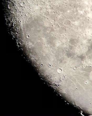
India's Chandrayaan-3 mission is the first successful attempt to land on the moon’s southern region and has uncovered several noteworthy discoveries on the lunar south pole using laser-induced breakdown spectroscopy (LIBS) and other spectroscopic techniques.
Arzak Mohamed from Macquarie University in Australia broke down how she uses spectroscopy to analyze ancient manuscripts.

A recent study used portable X-ray fluorescence analysis (pXRF) and scanning electron microscopy-energy dispersive spectroscopy (SEM-EDS) to uncover new insights into Stonehenge’s Altar Stone.

In a recent study, researchers used portable X-ray fluorescence (pXRF) analysis to determine how pottery vessels uncovered at Saqqara degraded over thousands of years.

A team of scientists recently analyzed a collection of late Roman vessels discovered in the United Kingdom.

A new study presents a novel WDXRF approach in determining carbon, oxygen, hydrogen, and nitrogen content in coal.

A team of researchers has conducted a successful round-robin test using total reflection X-ray fluorescence (TXRF) to analyze the elemental composition of rat tissue samples. The preliminary results demonstrate the effectiveness of TXRF in accurately determining the elemental composition of mammalian tissue.

Researchers have utilized an integrated wavelength-dispersive and energy-dispersive X-ray fluorescence spectrometer technique to comprehensively analyze bromine, iodine, and other components in soil samples, demonstrating an innovative method for quantitative elemental analysis of complex matrix geological samples.