Inside the Reactor Loop
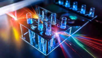
At 10:30 AM, Milos Burger of the University of Michigan presented "Laser Spectroscopy for Liquid Sodium Reactor Coolants: Design, Optimization, and Sensitivity Enhancement." His coauthors were Leandro Frigerio and Adam Burak (University of Michigan), Edward Kent (Argonne National Laboratory), and Joseph Craparo and Robert De Saro (Energy Research Company). The team built an immersion-compatible LIBS probe and testbed, then studied how probe design and operating conditions shaped signal quality. To improve oxygen sensitivity, they added two-photon absorption laser-induced fluorescence (TALIF) to the LIBS measurement. Sodium, potassium, and oxygen were all detected in liquid sodium, and tuning the excitation wavelength and inter-pulse timing improved the oxygen response. The work targets impurity monitoring in sodium-cooled Generation IV reactors.
Reading Carbon Through Plasma Timing
At 10:50 AM, Francisco A. Lopez-Linares of Chevron presented "Novel Time Resolved LIBS–Chemometric Approach for Comprehensive Carbon Analysis in Solids." Coauthors were Dayana Oropeza, Jose R. Chirinos, Xianglei Mao, and Vassilia Zorba (Lawrence Berkeley National Laboratory), Jinyi Han and Alexander Kuperman (Chevron), and Cesar F. Ovalles (CO2 Consulting & Services LLC). The group followed how CN and C₂ molecular emission evolved over time in plasmas formed in air from graphite, fullerene, nanotubes, and amorphous carbon, and used principal component analysis (PCA) to tell the samples apart. All carbon allotropes showed strong C₂ Swan band emission early in plasma formation (under 2 µs), while aromatic hydrocarbons emitted more strongly at later delays near 4 µs. The presenters suggested the approach could also estimate parameters of interest to the oil and gas industry, including H/C ratio, C=C bonding, enthalpy of formation, and total carbon content.
Lithium Straight From Brine
At 11:10 AM, Willis B. Jones of the University of North Florida presented "Direct Determination of Lithium in Brine Solutions Using a Liquid-Phase Laser-induced Breakdown Spectroscopy Instrument." Coauthors included University of North Florida undergraduates Micah X. DeCourcey, Abbey Gentile, Abigail J. Crossman, and Akane K. Bangay, as well as Jhanis J. Gonzalez and Chunyi Liu of Applied Spectra, Inc. The system introduced samples as a nebulized aerosol. Using a synthetic brine of 1% nitric acid saturated with sodium chloride, the team measured lithium at 670.8 nm with a detection limit of 0.3 mg/L. A comparison of external calibration, internal standardization, and standard addition showed that matrix effects strongly influenced accuracy, and standard addition allowed lithium quantification without dilution or further sample preparation.
Technetium Beside Its Parent
At 11:30 AM, John G. Lucchi of the University of Central Florida presented "Laser-induced breakdown spectroscopy of trace technetium in the presence of molybdenum: detection and quantitation." Coauthors were Zachary Murphy, Vasileios Anagnostopoulos, and Matthieu Baudelet (University of Central Florida), Mauro Martinez (Icahn School of Medicine at Mount Sinai), and project lead Hunter B. Andrews (Oak Ridge National Laboratory). Technetium matters both as a nuclear fuel cycle byproduct and as a medical radioisotope generated from molybdenum decay. The team immobilized technetium, together with molybdenum, in polymer films and surveyed technetium emission lines by LIBS. Using lines free of molybdenum interference, they reported a detection limit of 0.710 µg/mL and a quantification limit of 1.39 µg/mL.
Beating Intensity Clamping at a Distance
The session closed at 11:50 AM with Vassilia Zorba of Lawrence Berkeley National Laboratory and the University of California, Berkeley, presenting "Pushing the Boundaries of Ultrafast Laser Filamentation." Her coauthors were Sung-Uk Choi, Jose R. Chirinos, and Xianglei Mao (Lawrence Berkeley National Laboratory). Femtosecond filaments can carry laser energy over long distances for remote isotope detection, but intensity clamping caps the energy delivered to the target. Zorba described plasma gratings, formed when two or more synchronized filaments overlap and interfere, as a way around this limit. At equal energy, the gratings increased both overall emission and isotope signal compared with a single filament. The group also examined how inter-pulse angle and delay shaped the interaction with solid targets.
Infrared Photonics and Machine Learning: Making Vibrational Spectra Clinic-Ready
The BIM-Wed1: Machine and Deep Learning for Biomedical Diagnostics session met in Southern Pacific AG.
At 10:30 AM, Borislav Hinkov of Silicon Austria Labs presented "Infrared Photonics for Proactive and Predictive Healthcare," with coauthors Lukasz Sterczewski (Wroclaw University), Werner Mäntele (University of Frankfurt), and Johannes Kunsch (Laser Components). Drawing on a roadmap manuscript compiled by 85 researchers, Hinkov argued that mid-infrared (MIR) methods offer greater molecular specificity than near-infrared approaches because they probe fundamental vibrations.1 He described a shift toward reading the full spectral signature, or "IR-omics," enabled by Fourier transform infrared (FTIR) spectrometers, quantum and interband cascade lasers, and photonic integrated circuits. He also listed remaining hurdles: device performance, missing measurement standards, regulatory approval, and clinical trust.
At 10:50 AM, Oleg Ryabchykov of the Leibniz Institute of Photonic Technology (IPHT) presented "ML Refinement of Biomedical Vibrational Spectra," with Azadeh Mokari and Jhonatan Contreras (Friedrich Schiller University Jena) and project lead Thomas Bocklitz (Leibniz IPHT). Building on established spectral workflows, he discussed U-Net-style networks that suppress noise while preserving diagnostic features, two-dimensional spectral representations that tolerate wavenumber shifts across instruments, and Monte Carlo dropout to flag low-confidence predictions.2,3,4 Such tools, he suggested, could reduce the data needed per patient in clinical studies.
At 11:50 AM, Elizabeth A. Donkor of Oklahoma State University presented "Modified Alternating Least Squares Applied to Hyperspectral Imaging," with Thomas K. Hancewicz (TMH Associates), Karl S. Booksh (University of Delaware), Haoron Zhong (Guangxi Minzu University), and project lead Barry K. Lavine (Oklahoma State University). Modified alternating least squares (MALS) borrows from ridge regression to stabilize matrix inversion, allowing extraction of components near the noise level that conventional ALS misses. Coupled with adaptive baseline correction and library searching, MALS was applied to automotive paint forensics, polymer defects, disease detection, and meteorites.
Fluorescence EEM Spectroscopy: Fingerprinting Water and Biology
The afternoon MOL-Wed2: Recent Advances and Industrial Applications of Fluorescence Excitation-Emission-Matrix (EEMs) Spectroscopy session met in Southern Pacific B.
At 1:30 PM, Keith Gordon of the University of Otago presented "Fluorescence spectroscopy as a tool to characterize of New Zealand drinking water source variation over time." Using PCA and fluorescence regional integration, Gordon distinguished two water sources and tracked seasonal changes in algal, cyanobacterial, and humic-like signals. Welch's analysis of variance related the EEM data to turbidity, temperature, pH, conductivity, and colour. Algal and cyanobacterial contributions fell through the New Zealand autumn, consistent with greater activity in warmer, less acidic months.
At 1:50 PM, Jeffrey Julien of HORIBA Instruments presented "Beyond Single‑Mode Spectroscopy: Life Science Applications of A‑TEEM," with coauthor Lyufei Chen (HORIBA). A-TEEM collects UV–visible absorbance and full fluorescence EEMs in one simultaneous measurement. Julien outlined uses in characterizing complex biological systems, monitoring biochemical processes, detecting minor components in mixtures, and supporting multivariate analysis.
Summary
Whether the sample was a clinical specimen, liquid sodium, saturated brine, or a reservoir of drinking water, presenters treated deployment as the real test. Instrument advances, from immersion probes to interfering femtosecond filaments, were matched by attention to calibration, chemometrics, and machine learning. The remaining obstacles were stated openly: matrix effects, standardization across devices, and the clinical and regulatory trust that new methods must still earn.
References
(1) Hinkov, B.; et al. Infrared Photonics for Healthcare: A Roadmap for Proactive and Predictive Health Management. arXiv 2026, arXiv:2602.08248. https://arxiv.org/abs/2602.08248
(2) Storozhuk, D.; Ryabchykov, O.; Popp, J.; Bocklitz, T. RAMANMETRIX: A Delightful Way to Analyze Raman Spectra. arXiv 2022, arXiv:2201.07586. https://arxiv.org/abs/2201.07586
(3) Mokari, A.; et al. A Comparative Study of Robustness to Noise and Interpretability in U-Net-Based Denoising of Raman Spectra. Spectrochim. Acta, Part A 2025, 126577. https://www.sciencedirect.com/science/article/pii/S1386142525008844
(4) Contreras, J.; et al. ATR-FTIR Spectroscopy of Saliva and Machine Learning as a Screening Test for Sjögren Disease. Anal. Chem. 2025, 97 (47), 26034–26044. DOI: 10.1021/acs.analchem.5c04238.