
Mammograms can find breast microcalcifications but can’t tell whether they are likely benign or malignant.
Jürgen Popp, winner of this year’s Ellis R. Lippincott Award, discusses the regulatory, cost, and workflow hurdles of moving Raman spectroscopy into hospitals.
First of all, I'm completely excited because this is something that is really going close to my heart, and it's showing that with the research we are doing, we can really make changes in terms of medical diagnostics.
And I also have to say that this is an award for an entire team of people working on this with me for more than two decades. Many PhD students have done their work in this field. On the other side, many technicians and engineers have also been contributing to this research. And last but not least, all the medical people we worked together with. So there’s a lot of close collaboration.
The challenge is really multifaceted because it's not just about doing nice research in the laboratory. We start there, and we get samples from our clinical partners, and we are looking at the samples. It could be bacterial material, or it could be virus particles, or it could be tissue. Well, in the laboratory, a lot of things work quite nicely.
But if you then consider moving into the hospital, for example, into the Department of Clinical Chemistry, or if you want to go to clinical microbiology, or even if you want to go into the operation room, then things change completely because then you need hardware that is very solid that works the same way and where there’s no changes. In the lab, you can readjust everything quite easily, but if you are in a clinical environment, it just has to work.
On the other side, your software needs to be very reliable, and it should be very robust. Meanwhile, the technique used in the experiment should also be run by scientifically experienced personnel, but it should not be a chemist or a physicist working on this, so you also have to rethink the entire way of doing your experiments.
The next thing is that you have to translate the information into something that can then be easily understood by the clinicians because they don't like to look at the Raman spectrum. They want to have information, and this you also have to translate, so this is a multi-faceted challenge.
I'm quite happy that in Jena, we have an environment at the Leibniz Institute of Photonic Technology where we have all these opportunities to go from the lab to a prototype and maybe in the future also to a product. This needs a lot of experience in system technologies and software development. And then, on the other side, having the physicians who are very much interested in these new technologies. But you always have to think about the workflow.
There are also additional obstacles. We have in Germany now a new medical device regulation. If you want to do experiments on humans, you have to fulfill a lot of requirements. You have to do a lot of documentation to do a first experiment on a human. On one side, this is a pain in the neck. On the other side, since we know how this is working, this gives us an added value because now when we develop techniques, we know where we have to look. So in the beginning, it was a nightmare, but now I think it's really a benefit.
Regulation is one thing that is really challenging, but it's needed, because what you need at the very end is something trustworthy. Because we are using not just patient samples and we are working with humans directly, we need to have a technique which is really trustworthy.
The first thing is that you need to fulfill the medical device regulation, so you have to have something that is very robust and stable. You have to adapt the entire workflows. For example, if you talk about infection, you also need to fit into the workflow of clinical chemistry and clinical microbiology. If you don't fit, the technique will not be used; it will not be adapted. So, this is one of the challenges. You have to really consider and understand what is really needed.
The next challenge is that the technique should be better compared to the gold standard, but on the other side, it should also be cheaper because you should not add additional costs. There are a lot of great examples out there from industry. They are doing a very good job in terms of detecting infection-causing pathogens, looking at the resistance, but they don't really enter the market completely because they're way too expensive. This is also what we are considering.
So, we now really have two ways to look at infectious diseases. One is on the host response; one is directly looking at the pathogens themselves. For example, if we want to look at the pathogens and the antimicrobial resistance, then our test needs to be very cheap. This means we have to have a device that is low cost, which should be around 5,000 to 10,000 euros for measuring, but the test itself should be around a euro or less. This is the challenging part. At the moment, we have something like a test which costs 10–15 euros, and we have to reduce it by a couple of factors so that, at the very end, it can be a commercial product. It should be one or two cents in production.
For the host response, this is a test that does not exist. If we are successful, then this can change the entire setting in terms of how to treat patients. Because what we learn from our medical doctors is that if a patient comes with symptoms of a severe infection like severe sepsis into the emergency department, then there will be treatment with antifungal, antibacterial, or antimycotic medications because they don't know what is going on, so you are using a lot of medication at the same time. This is not really helpful, and it could also harm the patient, but this is what they have to do. If we could provide a test which already gives information in something like 10–15 minutes, then they already have better insights into how to treat the patient.
This is something we are trying to get into the market. But again, it's a medical device. You have to fulfill all these questionnaires, and this is a long way to go, but we are convinced that we can achieve this goal.
We have a really good collaboration with the head and neck hospital in Jena, and the director is really keen on using this new technique.
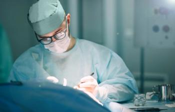
So, what does it mean when we do experiments and diagnostics in the surgery room? First, before the operation starts, our team comes, turns on the system and calibrates it, so when the surgeon is coming, everything is ready. This is the way we started. Then, when the surgeon comes, the patient is inside the operation room, and they do their classical tests, take the biopsies, and remove the tumor. In order to see where the tumor border is, for example, they use our Raman probe.
What do they need to learn? Well, what does a good Raman spectrum look like? This is something they have to learn at the moment. In the future, we can use some software tools to control this as well, but they have to know how to use the probe because with Raman itself and the devices, you can't really work when there is a lot of light around. Therefore, they need to consider how to measure so that we don't have the stray light from outside. Second, they have to know how to use the probe so that we get a nice Raman spectrum. And honestly, they learn it in a very short period of time. At the very beginning, they needed something like 10 minutes to get a good Raman spectrum. Now they can do it in one minute, so they can use it without really changing the workflow, and this is quite important.
So, the workflow may be changed in the future, but only in terms of the frozen section biopsy not being taken anymore then because at the moment, what they do is they remove the tumor and then take biopsies around the tumor to see whether there is still a tumor or not. And what they can do in a hospital like in Jena is frozen section analysis, but this is something which takes at least 30 minutes, and the accuracy is not as high.
This means they can also oversee a lot. Then, these biopsies are taken to the pathology laboratory, and very often they realize, "Oh, we have overseen something," and so the patient has to come back. With our Raman probe, this will be the only change: frozen section biopsies will not be taken. The classical biopsies will be taken, but based on the Raman spectra, the surgeon can make the decision where to cut and where he or she can leave the tissue inside.
What does it mean? We adapt ourselves exactly to the workflow, and then it can be easily used. I think this is quite important for all optical techniques. If we change, or if the requirement is to change, the entire workflow will mainly not be adapted. Therefore, you need to look at the workflow, you need to know how this is being done, and then you can really work inside the clinic.
And finally, you need a device which can then also be sterilized, for example, because this is a requirement. Our Raman probes are not single use, so you have to reuse them. Then, you also have to consider what is needed in order to fulfill these legal requirements. This was also quite challenging for our team because you have to prepare an optical probe that is going into the sterilization chamber. Then, the temperature is hot, and there are some chemicals and it should survive since we developed the probes in such a way that they can survive.
I think this concept is starting to get popular all over the world because what we realize is, at the moment, our healthcare system is organized in an episodic way. What does it mean? We go to the physician when we feel sick, and then we get our medication or, if it's needed, an operation, depending on our disease. But in between these different episodes, there is no information.
So now, with these new tools, especially wearables like Apple watches, we can already measure a lot of physical parameters. Even with some other variables, we can also measure our blood pressure. This means we get a lot of physiological information that we can monitor over time. This means, for example, we can easily monitor how our pulse and how the oxygenation level of our blood is developing over time. We can see whether there is something going on because we're doing exercises or we are sick.
What is missing, more or less, is the molecular information because a lot of things are really present in body fluids, cells, and tissue. If we can get this molecular information, we can add a new dimension. This means if we can continuously monitor, for example, some biomarkers. Very popular biomarkers would be glucose, or lactose if you're doing exercises, but also some inflammation markers. So, if you can, for example, maybe take a drop of blood, and this in a home care setting, and this maybe every day, a small drop of blood, and we measure our white blood count. Because with optical spectroscopy, we are not just looking at the white blood count itself. We're also looking at changes in the molecular pattern. This means maybe our baseline is changing, and then we can get an understanding of what is going on or at least see that a change is going on.
It's not that the AI tools should produce a lot of false positives, so that we go to the physician and we are not really sick, but it should come up with a good understanding of what the baseline is. If there is a change in the molecular markers, the physiological markers, then there should be an indication of whether you now have to go to a physician. This may lead to a situation where we can get into early detection, and this also means that we can go more to prevention. This is the concept of a digital twin.
Here at the conference, people are talking about optical twins. They are talking about digital twins, and when you talk, for example, also to physicians, they already want to have a digital twin or an organ. I think there is now an understanding that this might be very helpful, and it's more or less also coming from industry already. In industry, the digital twin is already quite common to look at the
Therefore, since the concept has already been considered as something helpful, many scientists have also realized this, and they are looking into miniaturization of optical devices. I'm pretty sure we can add this to the healthcare system, but there are certain requirements. The technique needs to be driven, and it needs to be developed, but this is just one step. It also needs to be implemented, and this means we have to talk to all the different stakeholders. It's about regulation because we need to have devices that are working and trustworthy. It's about the software; we need trustworthy AI. We need some cloud-based systems, and this needs to be built up because not everybody can build their own cloud-based system. It needs to be a big initiative from a society. In Europe, we have, for example, this virtual health twins initiative, and we hope that we can convince them that we can add this molecular information here to their twin initiative. We also need those initiatives worldwide.
Therefore, we should not just look at the technique itself, but we have to look at the entire ecosystem. If we do this nicely and correctly, then I would say in 10 years we can really have this already implemented.
This interview was edited lightly for clarity.