
A recent study out of Russia introduced a new method for identifying plant-based oils and adulterated dairy products.
A recent study out of Russia introduced a new method for identifying plant-based oils and adulterated dairy products.

Top articles published this week include highlights from the recently released “The Future of Forensic Analysis,” articles about detecting olive oil fraud, and an announcement from 3M regarding the winner of their Young Scientist competition.
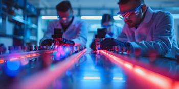
A compilation of recent studies that used laser-induced breakdown spectroscopy (LIBS) as part of the experimental procedure are presented.

With the release of Part 2 of “The Future of Forensic Analysis,” we break down what readers can expect from this issue.

Scientists from the University of Tokyo explored the utility of laser-induced breakdown spectroscopy (LIBS) in lunar missions.

NASA’s Perseverance rover is collecting valuable information for scientists back on Earth. A recent study explored how laser-induced breakdown spectroscopy (LIBS) is being used to analyze the Martian surface.

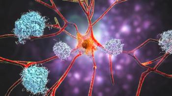
A recent study presents a new technique that combines femtosecond double-pulse laser-induced breakdown spectroscopy (fs-DP-LIBS) with machine learning (ML) algorithms to significantly enhance tissue discrimination and signal quality, paving the way for more precise biomedical diagnostics.
Sirish Subash is the winner of the Young Scientist Award, presented by 3M and Discovery education. His work incorporates spectrophotometry, a nondestructive method that measures the light of various wavelengths that is reflected off fruits and vegetables.

Researchers at Henan Agricultural University have developed a multi-channel magnetic flow device combined with surface-enhanced Raman spectroscopy (SERS) for the rapid and precise isolation, identification, and quantification of lactic acid bacteria and yeast, revolutionizing quality control in fermented food production.
Top articles published this week include an interview with Landulfo Silveira Jr., an article about using Raman spectroscopy in hematology, and a recap of a recent study that used infrared (IR) spectroscopy to screen for cancer.

In this paper, a system based on laser induced breakdown spectroscopy (LIBS) and back propagation (BP) method was developed for the composition and traceability analysis of crop burning smoke.

Researchers at Bilkent University and Sabancı University, led by Burak Ülgüt, have advanced the understanding of charge transfer processes in lithium batteries by employing electrochemical impedance spectroscopy (EIS) with varied parameters, revealing critical insights into battery performance and kinetics.

Researchers from Sichuan University and the University of Georgia have developed an advanced method combining Raman spectroscopy and chemometric analysis to effectively identify and distinguish between various PFAS compounds, improving detection and environmental monitoring capabilities.
A recent study published in Frontiers in Aging Neuroscience by Wenyu Jiang and colleagues in China found that patients with mild cognitive impairment (MCI) exhibit abnormal functional connectivity in the right prefrontal cortex as revealed by fNIRS, highlighting potential cognitive implications and the protective role of education.
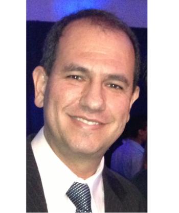
Spectroscopy sat down with Landulfo Silveira Jr. of Universidade Anhembi Morumbi-UAM and Center for Innovation, Technology and Education-CITÉ (São Paulo, Brazil) to talk about his team’s latest research using Raman spectroscopy to detect biomarkers of cancer in canine sera.
Top articles published this week include a SciX interview about mentorship, a feature article on wildlife crime, and a news article highlighting a new near-infrared (NIR) transient absorption spectrometer.

A recent study looked at how deep–UV Raman technology is studying the volcanic history of Mars.

A recent study examined the role of infrared (IR) spectroscopy in volcanology.

Here, a recap of a recent study that used Fourier transform infrared (FT-IR) imaging to evaluate changes in the inulin, lignin, and suberin contents of tuberous roots is presented.
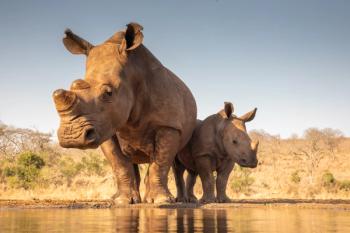
The emergence of new spectroscopic technologies has allowed investigators to solve and prosecute wildlife crimes more quickly.

This new study examines how spectroscopic techniques, such as attenuated total reflection Fourier transform infrared spectroscopy (ATR FT-IR), ultraviolet–visible–near-infrared (UV-Vis-NIR) spectroscopy, and Raman spectroscopy, were used to analyze the pigments in ancient Chinese wall paintings.

Here, we present a compilation of some of the most recent studies that used Raman spectroscopy as part of their methodology.
Top articles published this week include insights into machine learning and chemometrics presented at the SciX Conference, a compilation of recent studies in environmental analysis, and a news article highlighting the latest research in mineral classification.
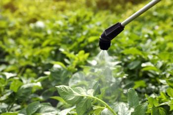
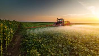
A recent study examined using surface-enhanced Raman spectroscopy (SERS) imaging in pesticide residue detection.

A recent study used spectroscopic techniques to study the mineral composition of sedimentary rocks in Texas.

Researchers in China proposed a new method using laser-induced breakdown spectroscopy (LIBS) in deep-sea mineral detection.

Five invited speakers joined Joseph Smith, the 2024 Emerging Leader in Molecular Spectroscopy, on stage to speak about trends in hyperspectral imaging, FT-IR, surface enhanced Raman spectroscopy (SERS), and more during the conference in Raleigh.

A compilation of some of the latest studies in environmental analysis is presented below.

This new study examined food contact materials (FCMs) and how mass spectrometric techniques have been used to measure harmful substances from FCMs that end up in food.
During the conference in Raleigh, North Carolina, experts from universities around the world spoke about their research using Raman, infrared spectroscopy, and more for biomedical imaging.