Researchers have found that LIBS spectroscopy models trained on compressed rock pellets produce inaccurate compositional readings when applied to fine-grained loose powders, raising concerns for planetary missions analyzing Martian soils.
Researchers have found that LIBS spectroscopy models trained on compressed rock pellets produce inaccurate compositional readings when applied to fine-grained loose powders, raising concerns for planetary missions analyzing Martian soils.

Abstract submissions are open through April 15 as conference expands scope to include molecular methods.
Metrohm's new i-Raman Platform is designed for both researchers and industry professionals alike. This Q&A overview provides the necessary information users would want to know.

Pittcon 2027 will take place in Pittsburgh, Pennsylvania, returning to where the conference was first held back in 1950. What does this mean for the Steel City?

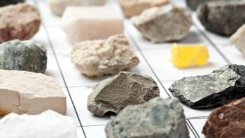
This partnership will digitize spectra from more than 120,000 mineral specimens, opening reference data to researchers across geology, forensics, materials science, and related fields.

A biennial United Kingdom conference returns with an expanded technical program, featuring artificial intelligence (AI) and biomedical imaging sessions alongside dedicated support for early-career scientists.

In this Q&A overview, we explore how these scientific advancements are reshaping our understanding of Ancient Egyptian history and culture.

In this review article, the authors highlight the latest advancements in infrared (IR) spectroscopy in gas drilling.

This overview of nearfield spectroscopy highlights how this technique operates and when it should be used.
In a press release, Metrohm USA announced that they will officially open a new, company-owned facility in Houston on March 24, 2026.

A recent study used minimally invasive synchrotron and spectroscopic techniques to fully characterize the materials, degradation processes, and conservation needs of a 26th Dynasty Ptah–Sokar–Osiris wooden statuette from Giza, establishing technical baselines for Saite workshop practices and future preservation of polychrome artifacts.

Interested in learning more about ultraviolet–visible (UV-vis) spectroscopy at Pittcon? The Spectroscopy editorial staff has you covered!
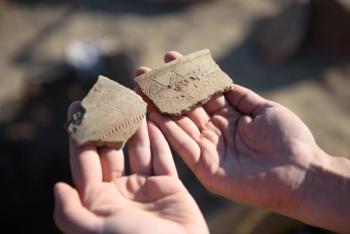
A continuation of our recap of a recent study published in Microchemical Journal highlights the implications of how Raman spectroscopy can help analyze ancient DNA remains.

A recent study shows that non-destructive Raman spectroscopy measurements of protein-to-mineral ratios in ancient teeth can accurately predict endogenous DNA preservation, enabling archaeologists to pre-screen specimens and avoid unnecessary destructive sampling.
This brief tutorial offers an overview of Raman spectroscopy and the scientist responsible for discovering this technique.

Top articles published this week include a couple new “Pathways in Spectroscopy” episodes, an interview with JAAS Prize and Nu Emerging Pioneer award winner David Clases, and a blog post that explores how early scientific ambitions translate to building a career in spectroscopy.

Researchers at Jiangsu University of Science and Technology showed that FT-IR spectroscopy combined with optimized chemometric modeling can rapidly and accurately detect and stage Bombyx mori nucleopolyhedrovirus infection in silkworms.

In a recent press release, CEA-Leti announced that a team of researchers from their institution and CEA-IRIG validated what they describe as the first chip-scale, battery-operated electron paramagnetic resonance (EPR) spectrometer.

A recent review article explores how machine learning (ML)-assisted Raman spectral classification is being used in applications such as biomedicine and material analysis.
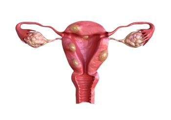
A recent study presented an approach combining Fourier transform infrared (FT-IR) imaging spectroscopy, histology, and statistical analysis that can identify biochemical spectral markers and distinguish benign from malignant uterine smooth muscle tumors.
Top articles published this week include the latest “Focus on Quality” column, an interview about handheld X-ray fluorescence (XRF) instrumentation, and the latest episode in “Pathways in Spectroscopy.”

A recent study demonstrates that updated predictive models based on NIR spectra can outperform traditional nitrogen-based prescreening methods in identifying samples suitable for radiocarbon dating.

In this Pittcon preview, we highlight some important talks, workshops, and symposiums happening during the conference.

A 2026 study published in the journal Remote Sensing tested Germany’s EnMAP hyperspectral satellite to see if it could identify and map acid mine drainage (AMD) and associated mineral residues from a legacy sulfide mine in Sardinia, Italy.
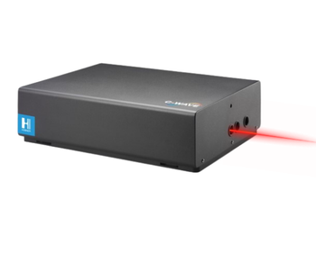
HÜBNER Photonics has announced the launch of the C-WAVE BTS, which is a continuous-wave (CW), single-frequency titanium:sapphire laser.

Validated green ICP-OES/ICP-MS workflow quantifies 25 toxic metals in wastewater at trace levels, guiding labs on tiered monitoring and compliance.

A recent study demonstrated that Fourier transform infrared (FT-IR) spectroscopy combined with chemometric modeling can accurately quantify ethanol in beer.

In this article, we highlight some of the important talks on atomic spectroscopy that will take place at Pittcon on Sunday March 8th.
Top articles published this week include a look at the top 10 applications of near-infrared (NIR) spectroscopy in biopharmaceutical analysis, an interview about the current trends in spectroscopy, and an inside look at handheld X-ray fluorescence (XRF) instrumentation.

Spectroscopists are routinely embracing complex algorithms to handle high-dimensional, nonlinear, and multimodal data. Why is this happening? We explore this question in this tutorial.