


This article discusses how FT-IR and SERS is being used to detect counterfeit pharmaceutical drugs.
This article discusses how FT-IR and SERS is being used to detect counterfeit pharmaceutical drugs.

This article discusses coherent Raman imaging and how it can visualize and quantify cutaneous pharmacokinetics (PK).

Working at the frontiers of biotechnology, fiberoptics, lasers technique, and molecular spectroscopy, Tuan Vo-Dinh of Duke University has developed multiple sensor technologies for medical research and diagnostics. Throughout this work, Vo-Dinh and his research colleagues have brought spectroscopy to biomedical applications. In this second recent interview, Vo-Dinh talks about his research work and philosophy.

An increasing number of antibiotic residue problems in food have emerged around the world. We examine how SERS is used to identify antibiotic residues in chicken, focusing on doxycycline hydrochloride and tylosin.

This year’s molecular spectroscopy award recipient is Bhavya Sharma, who is demonstrating research leadership focused on neurochemical detection using SERS and SORS Raman spectroscopy.

To better understand Raman spectral profiles, we briefly examine spectral line shapes, while discussing spectral shape, bandwidth, and broadening as percentage of Gaussian and Lorentzian components.

This article explains the key steps of using Raman technology to investigate carbon and carbon-based materials—such as carbon nanotubes, graphene, and carbon fibers and composites—as well as the process of analyzing the spectra.

In SERS experiments, the excitation laser power needs to be kept at a level that enables detection but avoids damaging the sample, as these examples illustrate.

Igor K. Lednev at the University at Albany SUNY in New York explains advances in forensic analysis using a variety of chemometrics techniques to classify ATR FT-IR and Raman spectra of bodily fluids.

Inline FT-NIR and offline terahertz Raman imaging analysis are used to characterize active pharmaceutical ingredient (API) crystallinity and to monitor different solid physical states of the API, to control process parameters of hot melt extrusion.

Using confocal Raman imaging and other advanced measurement techniques, we study the localized strain characteristics of tungsten diselenide (WSe2), an important nanomaterial used for optoelectronic device applications.
Using Raman imaging, wild-type and engineered yeast cells were compared for their ability to produce bioactive compounds. Raman imaging microscopy is able to visualize locales, relative abundance, and production efficiencies of biologically active compounds for the individual yeast cells.

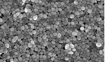
SERS can amplify Raman signals, but to make the technique practical for industrial use, large quantities of substrate are needed. The approach described here could enable cost-effective, reproducible manufacturing of SERS substrates at large scale.

Perovskites are known to be useful for fabrication of solar cells, and their crystalline structure plays an important role in their electronic properties. Here, we show how Raman analysis is able to confirm the presence of the required crystalline phase for solar cell production.
We illustrate how matching the optical properties of the microscope objective with the sample properties improves the spatial resolution and chemical speciation in depth profile measurements using confocal Raman microscopy.

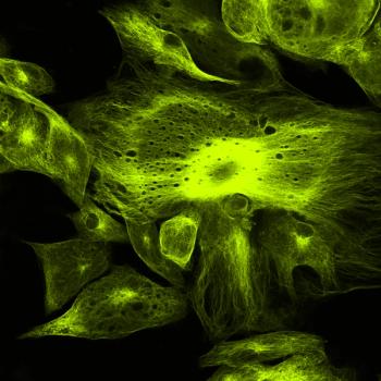
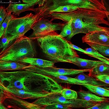
Raman spectroscopy offers promise for the evaluation of breast cell lines and tissue. Rina Tannenbaum of Stony Brook University discusses this work.

Fran Adar is the winner of the 2021 Gold Medal Award from the New York and New Jersey Society of Applied Spectroscopy (SAS).

Plasma spray–deposited metal films are used in many industrial applications. This study shows how high resolution terahertz time-domain spectroscopy (THz-TDS) can be used to analyze and characterize such films.

Surface-enhanced Raman spectroscopy (SERS), using gold nanoparticles, is useful for detection of low-levels of many analytes, including the water pollutant malachite green (MG).
SERS can be used for the detection and monitoring of drugs as pure compounds and mixtures. A demonstration of a sample preparation method used to detect components with weak substrate adsorption in the spectrum of a mixed solution is shown.

The application of data mining combined with data fusion of Raman and mid- infrared spectra was studied to improve discrimination ability for modeling the geographical origins of rice.
Raman and photoluminescence spectroscopy were combined with imaging to examine the spatial variation of solid-state structure and electronic character of two-dimensional (2-D) tungsten disulfide (WS2) crystals, which represent a family of new inorganic 2-D materials.

Raman measurements of chromite minerals demonstrated that chromium content could be accurately determined, supporting a possible application of portable Raman devices on Earth or in space for mineral analysis of asteroids and planets.
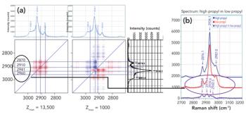
Raman spectra were measured in combination with 2D-COS analysis to understand how the addition of propyl side groups to a biopolymer backbone influences the structure of the polymer at the atomic level.

Zeolites are the most-used catalyst in industry. Synthesizing tailor-made zeolites is hampered by a poor understanding of how zeolite crystals actually form in solution. Scott M. Auerbach of the University of Massachusetts at Amherst is addressing this challenge with Raman spectroscopy.