


Raman spectroscopy offers promise for the evaluation of breast cell lines and tissue. Rina Tannenbaum of Stony Brook University discusses this work.
Raman spectroscopy offers promise for the evaluation of breast cell lines and tissue. Rina Tannenbaum of Stony Brook University discusses this work.

Fran Adar is the winner of the 2021 Gold Medal Award from the New York and New Jersey Society of Applied Spectroscopy (SAS).

Plasma spray–deposited metal films are used in many industrial applications. This study shows how high resolution terahertz time-domain spectroscopy (THz-TDS) can be used to analyze and characterize such films.

Surface-enhanced Raman spectroscopy (SERS), using gold nanoparticles, is useful for detection of low-levels of many analytes, including the water pollutant malachite green (MG).
SERS can be used for the detection and monitoring of drugs as pure compounds and mixtures. A demonstration of a sample preparation method used to detect components with weak substrate adsorption in the spectrum of a mixed solution is shown.

The application of data mining combined with data fusion of Raman and mid- infrared spectra was studied to improve discrimination ability for modeling the geographical origins of rice.
Raman and photoluminescence spectroscopy were combined with imaging to examine the spatial variation of solid-state structure and electronic character of two-dimensional (2-D) tungsten disulfide (WS2) crystals, which represent a family of new inorganic 2-D materials.

Raman measurements of chromite minerals demonstrated that chromium content could be accurately determined, supporting a possible application of portable Raman devices on Earth or in space for mineral analysis of asteroids and planets.
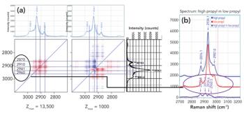
Raman spectra were measured in combination with 2D-COS analysis to understand how the addition of propyl side groups to a biopolymer backbone influences the structure of the polymer at the atomic level.

Zeolites are the most-used catalyst in industry. Synthesizing tailor-made zeolites is hampered by a poor understanding of how zeolite crystals actually form in solution. Scott M. Auerbach of the University of Massachusetts at Amherst is addressing this challenge with Raman spectroscopy.

Raman spectroscopy is extremely useful for characterizing crystalline materials.

Chemistry can help us understand the past.

The advantages of machine-learning methods have been widely explored in Raman spectroscopy analysis. In this study, a lightweight network model for mineral analysis based on Raman spectral feature visualization is proposed. The model, called the fire module convolutional neural network (FMCNN), was based on a convolutional neural network, and a fire-module was introduced to increase the width of the network, while also ensuring fewer trainable parameters in the network and reducing the model’s computational complexity. The visualization process is based on a deconvolution network, which maps the features of the middle layer back to the feature space. While fully exploring the features of the Raman spectral data, it also transparently displays the neural network feature extraction results. Experiments show that the classification accuracy of the model reaches 0.988. This method can accurately classify Raman spectra of minerals with less reliance on human participation. Combined with the analysis of the results of feature visualization, our method has high reliability and good application prospects in mineral classification.

Graphene exhibits special properties, such as high strength and high electrical and thermal conductivity and as such is highly desirable for key electronic components. A new Raman spectroscopy sampling technique has been applied to the characterization of batches of graphene that provides a simple, at-line method for obtaining key product data.

Raman spectroscopy is a valuable process analytical technology (PAT) for many applications across multiple industries, as a result of its many advantages, such as molecular specificity, ability to be directly coupled to a reaction vessel, and compatibility with solids, liquids, gases, and turbid media.

New Raman spectroscopy applications are emerging in non-traditional fields because of advances in easy-to-use commercial Raman spectroscopy instrumentation. With improvements in lasers, optics, and detectors, Raman spectroscopy has developed into a powerful measurement solution for manufacturing and quality control applications.

Spectroscopy Magazine spoke with Claudia Conti about her work in micro-SORS.

Spectroscopy Magazine sat down with Kay Sowoidnich to talk about how his group has demonstrated the potential of shifted-excitation Raman difference spectroscopy (SERDS) as an efficient tool for soil nutrient analysis.


SERS offers many advantages. However, there are several issues to be aware of when trying to use SERS signals in analytical applications.

Spectroscopy
In the past decades, we have witnessed the evolution of imaging technologies based on vibrational spectroscopy. In particular, the technical developments in Raman, coherent anti-Stokes Raman spectroscopy (CARS), and stimulated Raman scattering (SRS) microscopy allow researchers to gain new insights in biological, medical, and pharmaceutical studies.

Spectroscopy
Portable spectroscopic instruments have not had significant visibility within the scientific community compared with, for instance, the current generation of high-performance laboratory mass spectrometers.

Spectroscopy
Forensic traces are physical remnants of past events that provide critical information for criminal and civil investigations and adjudications. The scientific examination of traces is an incredibly valuable tool for forensic investigations, because the skilled interpretation of traces yields factual answers to a range of pertinent questions.
Special Issues
Click the title above to open the Raman Technology for Today's Spectroscopists 2020 special issue in an interactive PDF format.