


Webcasts
Webinar Date/Time: Thu, Aug 17, 2023 11:00 AM EDT

Life on Mars? Raman Spectroscopy Tests Stability of Biomolecules at Surface Conditions

The Detection of Life: How Spectroscopy Contributes to Space Exploration Missions
Webcasts
Webinar Date/Time: Thu, Aug 17, 2023 11:00 AM EDT

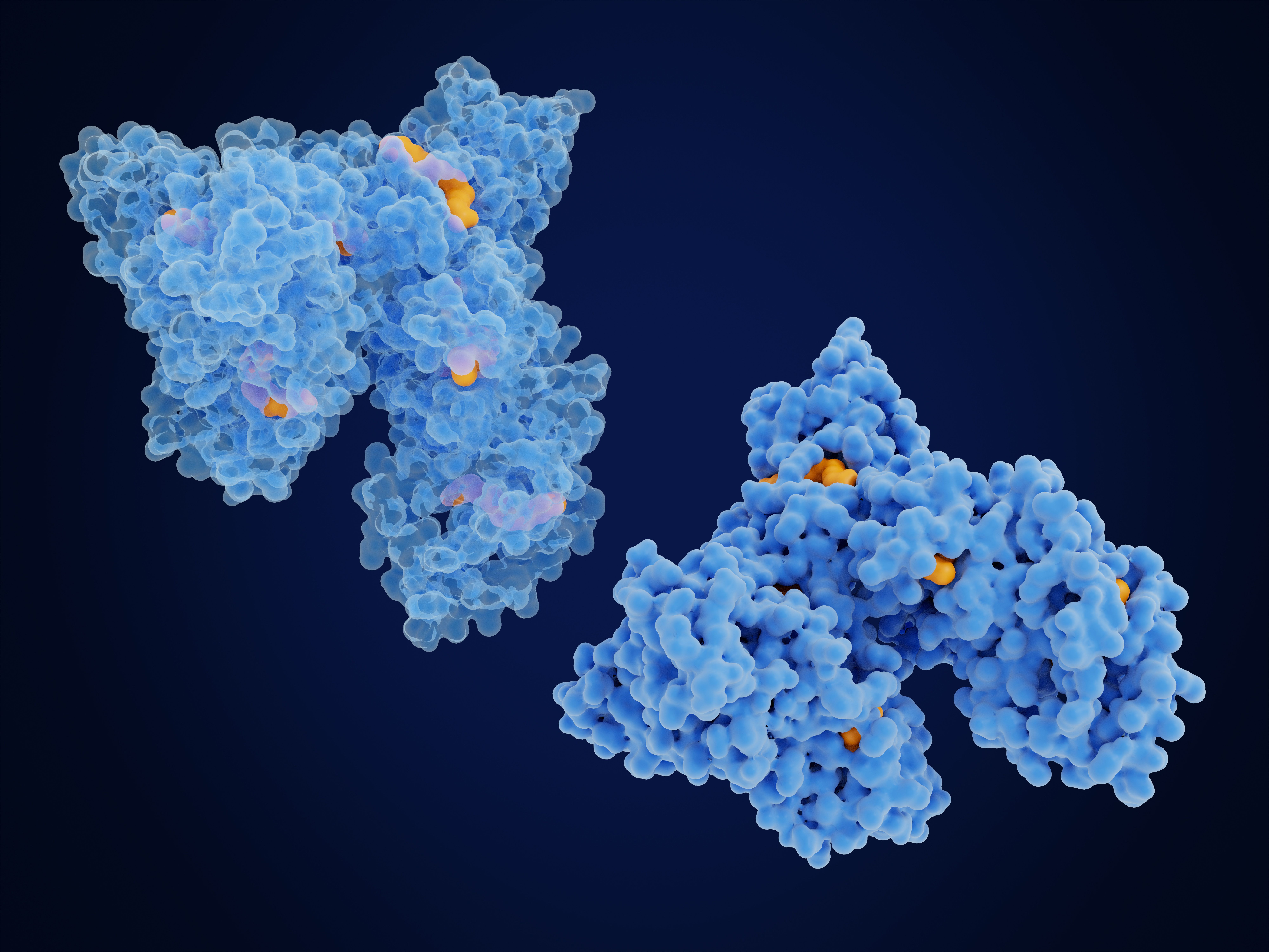
Researchers have developed a non-destructive method for identifying monoclonal antibody drug substances using Raman spectroscopy.

Webcasts
Webinar Date/Time: Tuesday, July 25th, 2023 Morning session: 9:30am EDT|6:30am PST|3:30pm CET|2:30pm GMT Afternoon session: 2pm EDT|11am PST|8pm CET|7pm GMT Wednesday,July 26th, 2023 Morning session: 9:30am EDT|6:30am PST|3:30pm CET|2:30pm GMT Afternoon session: 2pm EDT|11am PST|8pm CET|7pm GMT

In this interview, Jürgen Popp discusses the importance of Raman spectroscopy, where it can make a difference, and how it can be evolved and improved on in the future.
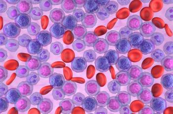
A new study reveals the potential of Raman spectroscopy in recognizing nucleophosmin (NPM1) mutant gene expression in leukemia cells.

Serum Raman spectroscopy combined with a convolutional neural network (CNN) offers a highly accurate and noninvasive method for diagnosing gastric, colon, rectal, and lung cancers paving the way for improved cancer screening and early detection.
A recent study reveals the pressure-induced phase transitions in imidazolium manganese-hypophosphite hybrid perovskite using Raman spectroscopy.

Researchers have conducted a preliminary study on the potential use of near-infrared (NIR) and Raman spectroscopy for predicting ice cream mix viscosity. The study highlights the promising performance of NIR spectroscopy and serves as a starting point for further investigations into in situ application of these analytical tools in the ice cream manufacturing process.

A new study demonstrates the improved accuracy of depth profiling in confocal Raman microscopy for analyzing the structural and chemical composition of polymeric microsphere layers.

Webcasts
Webinar Date/Time: Europe: Wednesday, June 28, 2023, at 2 pm BST | 3 pm CEST North America: Wednesday, June 28, 2023, at 1 pm PDT | 4 pm EDT Asia: Thursday, June 29, 2023, at 10:30am IST | 1 pm CST | 2 pm JST | 3 pm AEDT
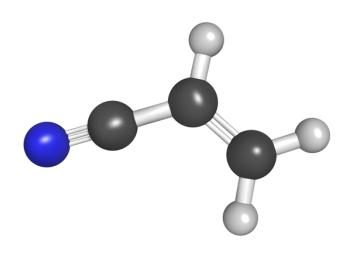
Raman spectroscopy has the capability of accurately determining the physical and chemical properties of bioplastics (polyhydroxybutyrate hexanoate [PHBHx]), which helps determine the maximum crystallinity a polymer can experience.

Double metal cyanide (DMC) can be monitored in real time using an in situ Raman spectroscopy method. In this study, real-time reaction progress information from in situ Raman results enabled researchers to accurately determine the reaction end point of DMC.

Determining the printing sequences of crossed writings and seal stamps is often difficult because the most common methods used are expensive, time-consuming, and cumbersome. A new method using Raman spectral area scanning offers a better alternative while conducting pigment analysis and determining intersection sequences of writings and seal stamps. We explain why.

Detecting illicit drugs in blood samples requires a rapid, non-invasive technique. The combination of surface-enhanced Raman spectroscopy (SERS) and chemometric techniques, such as principal component analysis (PCA) and partial least squares discriminant analysis (PLS-DA), can aid in this endeavor.

There is a growing desire among spectroscopists for having instruments small enough to be taken to the sample, as opposed to bringing the sample to the instrument. The result is that Raman spectrometers are becoming more miniaturized. Because these instruments come at a lower cost and offer distinct advantages over traditional spectrometers, the expectation is that a rapid expansion of when these instruments are applied will come forthwith. We offer a preview of how future miniaturized Raman spectrometers might look.

A recent study presents a detailed investigation on the pressure-dependent behavior of a Bi2(MoO4)3 crystal using Raman spectroscopy and lattice dynamic calculations. The study sheds light on the structural transformations and vibrational properties of the crystal under varying pressure conditions, offering valuable insights for material science research.
A new study utilized genetic algorithms to optimize pre-processing strategies and classification models such as SVM, multilayer perceptron, and PLS-DA for improved lung cancer diagnosis using Raman spectra data.

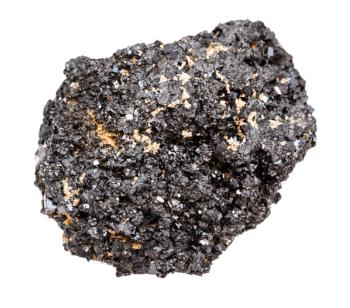
An in situ high-temperature Raman scattering study of monoclinic m-Ag2Mo2O7 microrods reveals an irreversible first-order structural phase transition and melting process, according to new research.

Scientists have investigated the structural changes of a discotic liquid crystal during its phase transitions using a combination of quantum chemical approaches and vibrational spectroscopy.

Biomedical Raman imaging is growing in the biomedical space, where technical advances and new information processing tools and techniques aim to propel the field into the future. An upcoming conference in Atlanta, Georgia, will explore these developments while bringing scientists in this field together.

The article discusses a comparison of Raman imaging assessment methods for phase determination and stress analysis of zirconium oxide layers and their application in the development of zirconium alloys, especially for nuclear applications.

This interview with Young Jong Lee highlights the work he and his team have done to reinvent solvent absorption compensation (SAC), and the potential it has across multiple forms of spectroscopy.

Researchers have developed a new method for analyzing Raman spectroscopy data in biological samples using group- and basis-restricted non-negative matrix factorization (GBR-NMF) framework, providing a promising approach for interpreting Raman spectroscopy data in biological samples.

Our annual review of new products for atomic and molecular spectroscopy, including details by category and highlights of overarching trends.

SERS of centrifuged blood serum samples of diabetic type II patients using 50 KDa filter devices can diagnose the disease at an early stage by studying small molecular weight proteins.