


A recent study from the Southern University of Science and Technology proposes a new Raman spectral preprocessing scheme based on self-supervised learning (RSPSSL).
A recent study from the Southern University of Science and Technology proposes a new Raman spectral preprocessing scheme based on self-supervised learning (RSPSSL).
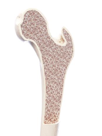
In a new study, scientists are investigating Raman spectroscopy as a technique for monitoring postmenopausual osteoporosis.

Scientists from Hirosaki University Graduate School of Science and Technology in Japan evaluated the eating quality of white rice samples using Raman spectroscopy.
Narangerel Altangerel, Zhenhuan Yi, and Marlan Scully of Texas A&M University recently used TRIP to analyze eight protein–ligand systems. Spectroscopy recently spoke to these three researchers about their findings and what the implications are for high-throughput drug screening.

Industrial scientists, in the spectroscopy community, scour the literature and scientific conferences to buy the technology they need.

Experts discuss Raman/SERS approaches across a number of different fields.

A recent study published in Advanced Photonics looks at three-dimensional (3D) imaging of cells and tissue using phase-modulated stimulated Raman scattering tomography (PM-SRST).

A team of scientists from the Masschusetts Institute of Technology are combining Raman spectroscopy with holo-tomography to monitor early embryonic development.

Here, we will show the chemical and spectral changes that occur during the cure of a commercial epoxy.

The field of analytical chemistry is well established. It’s time to develop the field of analytical biology—an emerging discipline that blends various research fields to provide a holistic view of biological phenomena.

Miri Park of the Fraunhofer Institute for Environmental, Safety, and Energy Technologies is examining how Raman spectroscopy could aid non-destructive sensing in agricultural science. Recently, Park sat down with Spectroscopy to discuss micro-Raman spectroscopy's role in assessing crop quality, particularly secondary metabolites, across different contexts (in vitro, in vivo, and in situ), while suggesting future research for broader application possibilities.

Dmitry Kurouski of Texas A&M University speaks to Spectroscopy Editor Patrick Lavery about Raman spectroscopy's role in determining crop yield of key food items as the world population continues to increase.

Spectroscopy spoke with researchers from the Columbia Climate School about how they are using stimulated Raman scattering microscopy to test for nanoplastics in water bottles.

This interview with Pola Goldberg Oppenheimer of the University of Birmingham highlights new research her team is working on that includes a Raman-based system for detecting early traumatic brain injuries.

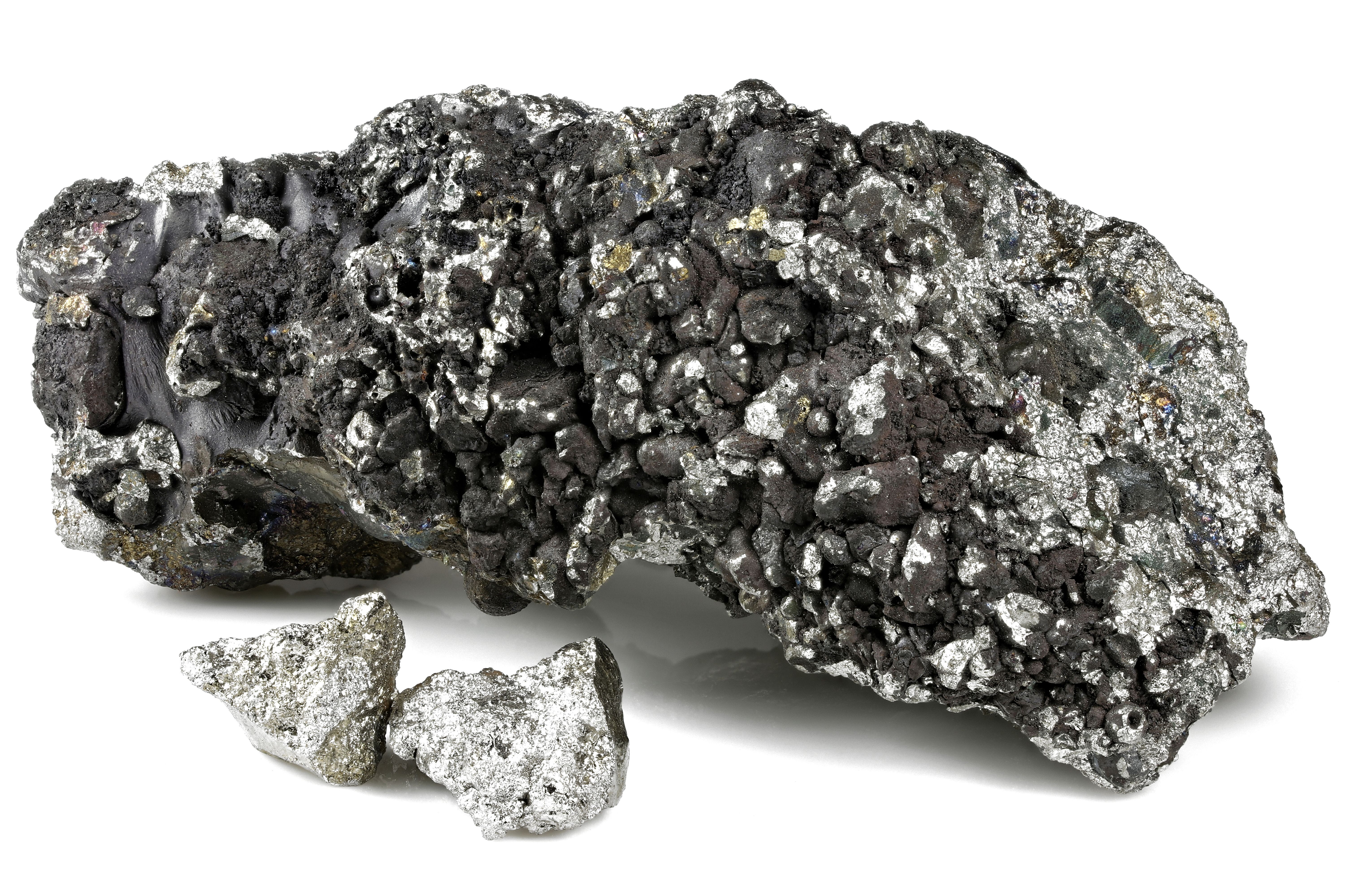
Triethylaminium picrate (TEAP) crystals were developed using a slow evaporation solution growth method and then characterized for their feasibility in optoelectronic and other uses.
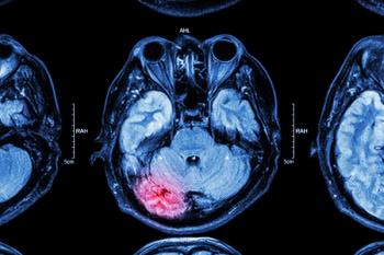
Scientists from the United Kingdom recently created a Raman spectroscopy-based system for detecting early signs of traumatic brain injury (TBI) in patients.

Webcast
Webinar Date/Time: Tue, Jan 23, 2024 11:00 AM EST
The editors of Spectroscopy retrospect on the most read articles of the past year.

A truly innovative approach to Raman spectral analysis is introduced in a newly published Applied Spectroscopy paper, utilizing a dynamic neural network modeling to significantly enhance baseline correction accuracy and offering a paradigm shift in spectral preprocessing.

The use of polarized Raman microscopy to complement the polarized light micrographs of spherulites in polymers reveals much about the orientation behavior of the lamellae in spherulites.

Researchers at NASA's Johnson Space Center, led by Ryan S. Jakubek, have unveiled a novel calibration method in Applied Spectroscopy that enhances the precision of Raman measurements conducted by the SHERLOC instrument on the Perseverance rover, providing a clearer and more accurate representation of intrinsic Raman spectral bandwidths on Mars.

Webcasts
Webinar Date/Time: Tuesday, January 9, 2024 at 11am EST | 8am PST | 4pm GMT| 5pm CET

Spectroscopy's laureate series will highlight the lives and careers of some of the most influential vibrational and atomic spectroscopists.
This study explores the enhanced performance of modified alternating least squares (MALS) over alternating least squares (ALS) in analyzing infrared and Raman image spectral data, highlighting the stability and computational efficiency of MALS.

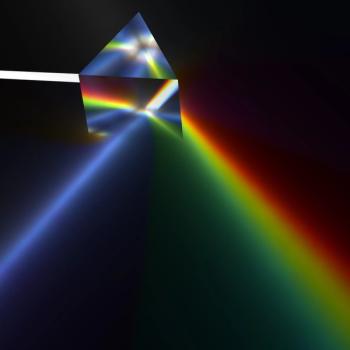
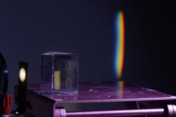
An inside look at the fundamentals of Raman microscopy and how Raman can be utilized in chemical imaging and analysis, from its inception to modern applications.