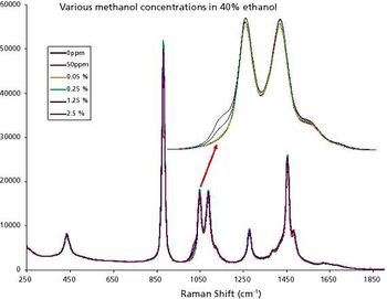
Application Notebook
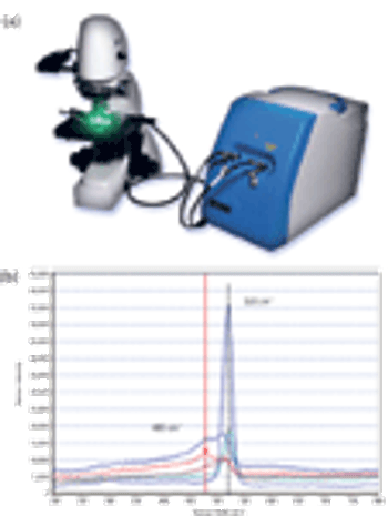
FTIR spectroscopy has long been used for the analysis of art and historical objects in support of efforts to conserve, restore, and validate authenticity of these rare objects. The value of the technique for this application lies in its inherent sensitivity, specificity, and non-destructive capabilities.