All News


DNA strand and Cancer Cell Oncology Research Concept 3D rendering | Image Credit: © catalin - stock.adobe.com

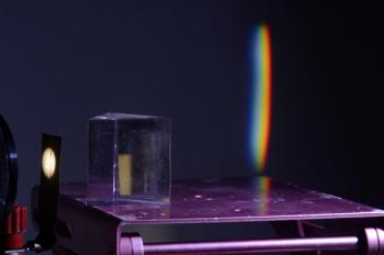
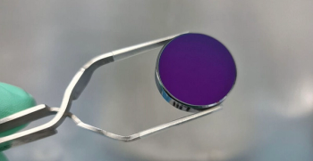
A recent study from Spain used surface-enhanced Raman spectroscopy (SERS) to study cancer cells with methylthioadenosine phosphorylase (MTAP) deletions, shedding new insights into the metabolic interactions inside the tumor microenvironment that could influence cancer aggression.

Small pile of minerals extracted in a rare earth mine. Ai generated. | Image Credit: © Joaquin Corbalan - stock.adobe.com.

The article describes a method for geochemical sample analysis using polytetrafluoroethylene (PTFE) crucibles and ICP-MS, achieving detection limits below 0.2 μg/g and relative standard deviations ranging from 1.1% to 16.4%. The approach minimizes acid usage, prevents dust contamination, accurately determines volatile elements, and is deemed suitable for high-throughput laboratories with numerous samples and diverse elements to be tested.

A one-inch-diameter silicon substrate coated with a conventionally deposited interference coating.| Image Credit: © Valentin Wittwer – Institut de Physique, Université de Neuchâtel, Switzerland.

Researchers from Thorlabs Crystalline Solutions, the University of Vienna, and the National Institute of Standards and Technology have achieved a revolutionary breakthrough, publishing in Nature Communications, with mid-infrared supermirrors exhibiting finesse exceeding 400,000, promising unprecedented sensitivity for applications in trace gas sensing and precision spectroscopy.

Extreme macro of polyester stable fiber on blue background | Image Credit: © Taigi - stock.adobe.com

Researchers at Kochi University and RIKEN have unveiled a new method for distinguishing individual polyester fibers in forensic investigations. Published in Spectrochimica Acta Part B: Atomic Spectroscopy, their advanced X-ray analysis refreshes how we unravel the composition of these fibers.

Messenger RNA or mRNA strand 3D rendering illustration with copy space. Genetics, science, medical research, genome replication concepts. | Image Credit: © Matthieu - stock.adobe.com

Duke University researchers, led by Joy Q. Li, revolutionize biomedical diagnostics with a multiplexed SERS-based nanosensor called inverse molecular sentinel (iMS) for micro-RNA detection, employing machine learning, particularly convolutional neural networks (CNN) and non-negative matrix factorization (NMF), to achieve higher accuracy in spectral unmixing, paving the way for more precise and efficient clinical diagnostics.