

Spectroscopy
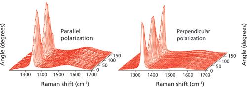
Polarization/orientation micro-Raman spectroscopy promises to be an important analytical tool to complement micro-X-ray diffraction.
Spectroscopy
Polarization/orientation micro-Raman spectroscopy promises to be an important analytical tool to complement micro-X-ray diffraction.
Spectroscopy
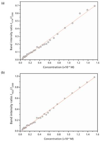
A quick and accurate method for detecting the antifungal agent crystal violet in aquaculture.
The Coblentz Society and the Federation of Analytical Chemistry and Spectroscopy Societies (FACSS) recently named Professor Duncan Graham of the University of Strathclyde (Glasgow, Scotland) as the recipient of the Coblentz Society?s 2012 Craver Award.
Spectroscopy
Raman has a unique capability to characterize nanoscale materials that are between crystalline and amorphous.
Spectroscopy
Confocal Raman microscopy can identify particles in the 5–50 ?m range and can bridge the gap between micro-FT-IR and SEM-EDS analyses.
Application Notebook
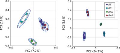
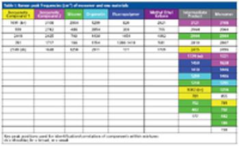
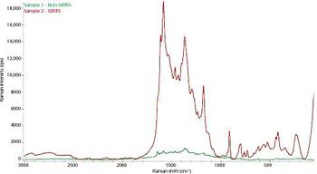
In the three decades since its discovery, surface-enhanced Raman scattering (SERS) has been used in numerous applications to increase signal intensity in Raman scattering experiments. The current study provides insight into the more practical aspects of enhanced Raman sampling for laboratory users. We describe how the signal enhancement from a surface-enhanced resonant Raman scattering (SERRS) process improves the ability to discriminate between ink samples using principal component clustering.
Spectroscopy
Temperature measurements can be made using spectral features such as the position, linewidth, and intensity of the Raman signal associated with specific optical phonon modes. Each of these spectral characteristics offers particular advantages, depending on the type of device and operational considerations.
Spectroscopy
A critical review focused on the Raman spectroscopy of carbonaceous materials and of polymer-based nanocomposites that contain carbonaceous (nano) materials as fillers
Spectroscopy
How can you navigate the maze of choices for detecting molecular vibrations with mid-infrared (IR), near IR (NIR), and visible (Raman)? Understanding what is being measured, how it is measured, and the advantages and disadvantages of each technique, will help.
Spectroscopy
In the three decades since its discovery, surface-enhanced Raman scattering (SERS) has been used in numerous applications to increase signal intensity in Raman scattering experiments. The current study provides insight into the more practical aspects of enhanced Raman sampling for laboratory users.

Spectroscopy
A few key steps will protect your samples and help ensure accurate results.
Spectroscopy
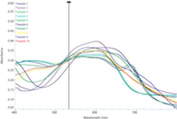
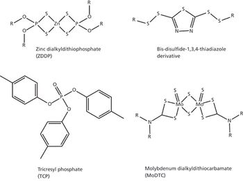
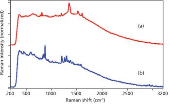
Developments in lasers, detectors, low-cost instruments, and fiber-optic probes have greatly expanded the lubrication systems being studied by Raman spectroscopy.
Spectroscopy
Surface plasmon resonance, charge-transfer resonance, and their combination determine the enhancement of surface-enhanced Raman scattering signals, and the varying intensities of the signal at different pH levels may result from the change in contributions of the combined system.
Special Issues
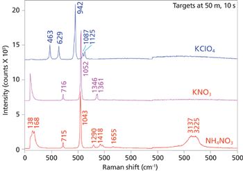
A portable Raman analyzer with laser excitation at 1550 nm provides "eye-safe" explosives detection.
Special Issues
A compact standoff Raman system can be used to detect hazardous chemicals and chemicals used in homemade explosives synthesis.
Spectroscopy
Spatially offset and transmission Raman spectroscopy enable chemical characterization of diffusely scattering samples at depths not accessible by conventional Raman methods.
Spectroscopy
Raman spectroscopy can be used to measure the vibrational spectra of both organic and inorganic materials.
Spectroscopy
Graphene has potential applications ranging from computer monitors to solar cells, and Raman spectroscopy is a useful method for its characterization.
Application Notebook
BaySpec, Inc. has developed a complete line of 1064 nm excitation, dispersive Raman systems that offer maximum reduction in fluorescence interference from biological samples and thus making them very useful tools for biofuel research.
Application Notebook
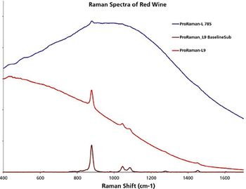
In recent years, the spectroscopy community has observed rapid development of Raman instrumentation and its usefulness in a variety of applications. Routine Raman analysis with 785 nm excitation has served well for the great majority of industrial applications and has become the most favored instrument configuration.

Spectroscopy
Virtually everything we know about stars is based on spectroscopy, including what we know about magnitude, red shift, and why the night sky is dark.
Spectroscopy
This month's column discusses the various multiphoton spectroscopy techniques and the lasers required for each approach.

Application Notebook
For the characterization of the properties of a sample with Raman spectroscopy, an ultrasensitive confocal Raman microscope allows the acquisition of a Raman image stack revealing 3-D information on the distribution of the chemical compounds.
Application Notebook
Low concentration natural methanol exists in most alcoholic beverages and usually causes no immediate health threat.
Application Notebook
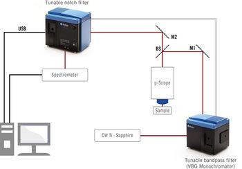
Photon etc. has designed two narrowband tunable filters for resonance Raman spectroscopy.